| |
|
|
INTRODUCTION Constrictive pericarditis. Pericardial
constriction is a chronic condition characterized by a thickened,
heavily fibrotic and inelastic, and often calcified, pericardium,
which leads to impaired diastolic filling of the heart. The term
constrictive pericarditis (CP) should be used when pericardial
constriction is associated with pericarditis [1]. Constrictive
pericarditis is a consequence of chronic inflammation of the
pericardium and is characterized by scarring, fibrosis and complete
loss of pericardial elasticity. The main symptoms are dyspnea, edema,
orthopnea and fatigue. The classic clinical picture is most often
characterized by isolated right-sided heart failure, with normal or
near-normal natriuretic peptide levels. Signs of venous congestion,
hepatomegaly, pleural effusions and ascites may occur [2,3,4].
Constrictive pericarditis should initially be treated with empirical
anti-inflammatory therapy for at least 3 to 6 months according to
the 2025 recommendations of the European Society of Cardiology
[1,5]. Patients in whom medical treatment has not led to
improvement, as well as patients with pericardial constriction not
associated with pericarditis, should be referred without delay for
cardiac surgical treatment, i.e. pericardiectomy [1,5]. Severe
hypotension may be a sign of chronic pericarditis with constriction
and cardiac tamponade (CTP – constrictive tamponade physiology),
which clinically manifests as Beck’s triad: hypotension, jugular
venous distension and muffled heart sounds.
Transient constrictive pericarditis. Transient
constriction is a form of constrictive pericarditis (CP) with active
inflammation present, which may resolve after anti-inflammatory
therapy or regress spontaneously. It develops during pericarditis
with or without pericardial effusion (effusive-constrictive
pericarditis). In more than 50% of cases, constriction resolves
after anti-inflammatory therapy [6,7,8]. In patients with newly
diagnosed constrictive pericarditis, a trial of anti-inflammatory
therapy lasting 3 to 6 months is recommended before referral for
pericardiectomy [9,10]. To demonstrate active pericardial
inflammation, the use of biomarkers (e.g. elevated C-reactive
protein) and multimodality imaging is necessary, including
cardiovascular magnetic resonance (CMR) [10,11].
Constrictive pathophysiology
It is important to distinguish constrictive pathophysiology from
actual symptoms of heart failure [3,6,7,12]. Echocardiographic and
hemodynamic signs of constriction may be present, such as: “septal
bounce” (paradoxical or abrupt movement of the interventricular
septum), respiratory phase-dependent shift of the interventricular
septum and Doppler features of constriction, while the patient may
be free of heart failure symptoms. The non-inflammatory phenotype of
pericarditis is a form whose pathogenesis, with normal C-reactive
protein levels, is unknown, and these cases are often
therapeutically challenging and more difficult to treat [13].
Treatment of constriction
The algorithm for the treatment of constriction uses clinical data,
biomarkers and imaging methods, including CMR (cardiovascular
magnetic resonance), CT (computed tomography) and ECG
(electrocardiogram) [1,5].
Empirical anti-inflammatory therapy is justified only in patients
with evidence of pericarditis based on clinical criteria, biomarkers
and/or imaging findings. Stratification of constrictive pericarditis
by response to therapy:
- Low risk: clinical remission after therapy, normalization
of imaging findings.
- Intermediate risk: improvement of symptoms without complete
remission, partial regression of signs of constriction.
- High risk: persistent symptoms, permanent signs of
constriction on imaging.
Constrictive pericarditis is a complex clinical syndrome
resulting from impaired diastolic filling of the cardiac ventricles
due to thickening, fibrosis, inflammatory changes or other
pathological changes of the pericardium. The clinical picture
depends on the degree and duration of constriction, and signs of
systemic venous congestion often dominate, including peripheral
edema, ascites, hepatomegaly, pleural effusions and exercise
intolerance. At the same time, left ventricular systolic function
may remain preserved, which can make differentiation from HFpEF and
restrictive cardiomyopathy difficult [1,14].
Echocardiography plays a central role in the initial recognition of
the constrictive physiological pattern. Of particular importance are
paradoxical movements of the interventricular septum, respiratory
variations in diastolic filling, the so-called annulus reversus: a
higher tissue Doppler velocity of medial e′ than of lateral e′, and
certain hepatic vein flow patterns. A combination of several
echocardiographic parameters increases diagnostic reliability, while
individual signs are not sufficient for a definitive diagnosis [13].
In patients with preserved ejection fraction, heart failure may
result from impaired diastolic function and elevated filling
pressures, whereby secondary pulmonary hypertension and right
ventricular failure may also play a significant role. Current
criteria for HFpEF therefore require, in addition to symptoms and
preserved ejection fraction, objective evidence of structural or
functional abnormalities and/or elevated filling pressures [16].
A particular diagnostic problem in this case is the simultaneous
presence of marked leukocytosis, hepatosplenomegaly and pathological
changes of the pleura and pericardium. In such a context, a
hematological process may play a significant role in the
differential diagnosis, including infiltrative and extramedullary
processes. However, the peripheral blood finding alone is not
sufficient to establish a definitive diagnosis of myeloproliferative
neoplasm [17-21].
The aim of this report is to present the complex clinical course of
a patient with severe systemic congestion, preserved left
ventricular systolic function, echocardiographic signs of suspected
constrictive pericardial physiology, pulmonary hypertension and a
concurrent finding of marked leukocytosis compatible with a
myeloproliferative process, as well as the diagnostic difficulties
due to the patient’s non-cooperation.
CASE REPORT:
MEDICAL HISTORY: We present a 73-year-old female patient with
long-standing cardiovascular problems: hypertension, mitral
regurgitation, pericardial adhesions and a small pericardial
effusion from 2014. Based on the documentation she brought, newly
developed G3 kidney disease is recorded; for 10 years she has had
type 2 diabetes mellitus on insulin therapy, as well as
hypothyroidism and marked leukocytosis. In May 2026, the patient was
hospitalized at the Internal Medicine Department of the Zaječar
Health Center due to a feeling of shortness of breath (dyspnea) and
received therapy from which she did not feel any significant
improvement. During the 2 months after this hospitalization in 2026,
a pronounced clinical picture of systemic venous congestion
developed, with orthopnea, bilateral pleural effusions, ascites,
peripheral edema and hepatosplenomegaly, and typical ANASARCA. On
13.07.2026, i.e. the day before admission to the "Dr Bastać"
Practice, she was discharged at her own request from the Internal
Medicine Department of the Zaječar Health Center.
MAIN COMPLAINTS are marked fatigue, inability to walk,
instability, episodes of fainting, a feeling of severe suffocation,
difficulty breathing, shortness of breath, occasional stabbing chest
pain and leg swelling.
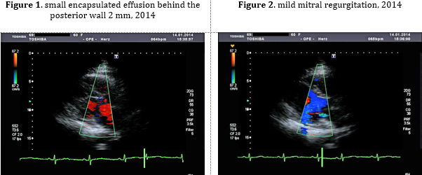
The first contact was back in 2014 - at that time, echocardiography
revealed mild mitral regurgitation, separation of the pericardial
layers, with adhesions (Figures 1 and 2) on the lateral wall - signs
of adhesive pericarditis, and a small encapsulated effusion behind
the posterior wall of 2 mm. Tissue Doppler showed that: systolic and
diastolic annular velocities were reduced, E/e’ ratio 10,6
borderline. In the laboratory findings, only thrombocytopenia
dominates, and since then she has been on bisoprolol 2,5 mg.
Thrombocytopenia.
Now, on admission, she gives no information and has no
documentation that she was hospitalized at the hematology department
of the Clinical Center Niš in 2024. It was later learned from the
heteroanamnesis that a bone marrow biopsy had been performed, but
the sample was not diagnostic. She refused the proposed further
treatment and was discharged with a diagnosis of splenomegaly and
suspected chronic myeloid leukemia. She also had laparoscopic
gallbladder surgery with complications and conversion to classic
laparotomy due to a calculus in the common bile duct.
CURRENT previous therapy: spironolactone 100mg 2x1, Bisoprolol
2,5mg 1x1, Empagliflozin 10mg 1x1, Ramipril- 5mg 1x1/2, Thyroxine
25mcg 2x1, Furosemide forte 500mg 1x1.
STATUS PRAESENS ON ADMISSION: 14.07.2026 – inspection
reveals extreme dyspnea, Tachypnea, distended neck veins, orthopnoea-
can lie down, examination difficult overall. Blood pressure was
130/70 mmHg, body weight 77 kg, and SpO2 95%. She refuses
hospitalization and referral to a tertiary institution (written
statement).
COR: HR 75 min, heart sounds muffled, rhythm regular, systolic
regurgitant murmur over Erb’s point 2/6. PULMO: breath sounds
diminished bilaterally at the bases, dullness to percussion up to
both scapular angles, early and late inspiratory crackles above the
dullness, signs of large bilateral pleural effusions (hydrothorax).
ABDOMEN: Soft and non-tender on palpation. The liver is palpable 4
cm below the RCM, the spleen is palpable up to 4 cm below the LCM.
EXTREMITIES: large cold doughy edema of the lower legs 3+,
circumference measurement: left leg 50/39/27cm, right leg
47/36/27cm, peripheral arterial pulses are symmetrically palpable.
LABORATORY ANALYSES ON ADMISSION 14.07.2026.
Urgent "point of care" laboratory analyses performed at the "Dr
Bastać" Internal Medicine Practice dominated by: very high
N-terminal pro-B-type natriuretic peptide (NT-proBNP 1175 pg/mL),
signs of renal dysfunction with creatinine 122 µmol/L and GFR 37 mL/min/1,73
m², as well as hyponatremia of 130 mmol/L. hyperuricemia: 774
µmol/L, albumin normal 37 g/L, leukocytes 55x109 /cm3, neutrophilia
84%.
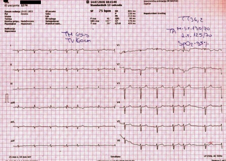
ECG ON ADMISSION: sinus rhythm with frequent supraventricular
extrasystoles SVES, HR 75/min, low voltage of R waves, no R
progression to v6 with deep S, extreme right axis with incomplete
right bundle branch block, QRS duration 90ms and QTc interval 420ms
normal, overall depolarization vector right-sided, rS in D1-2, aVL.
rsr in V1, no R progression to V6, deep S in V2-V6 and no ST
changes.
Figure 3. ECG on admission 14.07.2026.
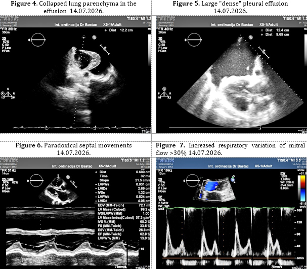
Ultrasonography of the pleura and lungs and
echocardiography on admission (technically difficult due to
orthopnea)
left - pleural effusion thickness approx. 9,7cm x 12,4cm x 7,6cm,
right – effusion approx. 5,3cm x 9,1cm x 4,4cm
Minimal ascites on the right between the liver and kidney 11 mm -12
mm, in the pouch of Douglas 9 mm (Figures 4 and 5).
Echocardiographic examination revealed a dilated left atrium,
moderate mitral regurgitation (MR), MR vena contracta 4mm, with
normal left ventricular dimensions. The interventricular septum
showed paradoxical movements - septal bounce shift, and preserved
left ventricular systolic function was recorded, (Teicholtz EF=76%,
and EF Biplane Simpson rule 65%, Figure 4). Diastolic dysfunction -
impaired relaxation and compliance - was also measured (E/e′=21,8,
E/A=2,0 and DTE=113ms) (Figure 5). The right ventricle was mildly
dilated, with TAPSE reduced to 12 mm and significant tricuspid
regurgitation TR 3+ and signs of moderate pulmonary hypertension:
elevated right ventricular systolic pressure RVSP=52 mmHg, The
pericardial layers were separated and moved in parallel, with
pericardial thickening: behind the posterior wall of the left
ventricle 7–8 mm-/ (Figures 6 and 7) / [12,2,13].
The next day, 15.07.2026, the patient was still orthopneic, 3 kg
lighter and with anasarca.
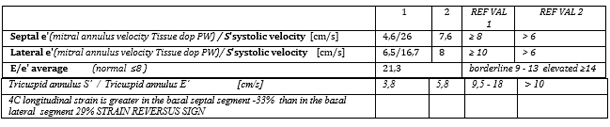
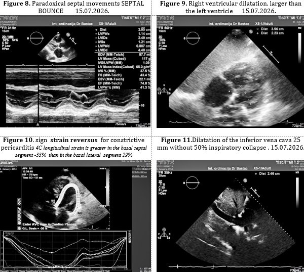
At that time the echocardiographic examination was supplemented
(Table 1). Echocardiography revealed preserved left ventricular
systolic function, EF 60 to 70%, paradoxical movements of the
interventricular septum, (Figures 6 and 8) and pericardial
thickening of 6–9 mm, parallel movement of the pericardial layers,
right ventricular dilatation and dysfunction, (Figure 9) significant
tricuspid regurgitation and pulmonary hypertension with an increase
in RVSP to 76 mmHg. The finding was interpreted as a syndrome of
global decompensated heart failure with preserved left ventricular
ejection fraction (HFpEF) with predominant right ventricular
failure, secondary pulmonary hypertension and signs of constrictive
pericardial pathophysiology - strain reversus (Figure 8) and
respiratory variation of Doppler flow greater than 25% (Figures 7
and 9). Constrictive pericarditis was suspected [1,13,14].
Table 1. supplemented echocardiographic
examination tISSUE DOPPLER AND STRAIN
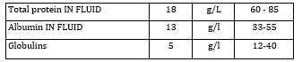
After both evacuating pleural punctures, a biochemical analysis
of the pleural fluid was performed 15.07.2026. clear transudate, and
at the same time abdominal ultrasound revealed splenomegaly, with a
spleen diameter of about 17–18 cm [4,17,18].
Table. 2. biochemical analysis of the pleural
fluid
COURSE AND THERAPY
Initial urgent parenteral therapy in the form of
a day hospital at the Int. practice of Dr Bastać
Due to the clinical picture of anasarca, ascites and bilateral
hydrothorax, intensive decongestive therapy was introduced,
including intravenous furosemide, spironolactone and an SGLT2
inhibitor, with further adjustment of therapy according to the
clinical condition [1,16].
Therapeutic course: Intensive decongestive therapy, supported by
bilateral therapeutic evacuating pleural punctures (500 ml and 350
ml), led to a turnaround in treatment and complete loss of dyspnea
on day 4 of treatment, with good regression of edema with high doses
of furosemide, spironolactone and empagliflozin, (with
antihypertensives paused during treatment within the day hospital
due to more severe episodes of hypotension), and was accompanied by
significant clinical improvement.
Key dynamics of therapy and of the therapeutic response to intensive
intravenous therapy (IV th) within the day hospital of the "DR
BASTAĆ" Internal Medicine Practice (Table 3).
Table 3. Overview of the clinical course and the
main intensive IV th within the day hospital of the "DR BASTAĆ"
Practice
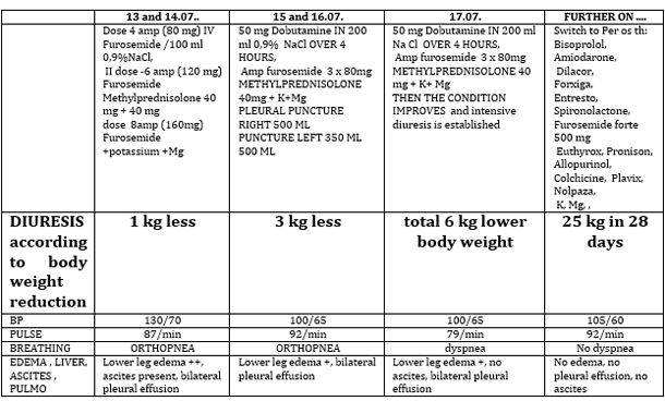
Over the following days, clinical improvement was
recorded with intensive diuresis and significant regression of
symptoms (Table 3).
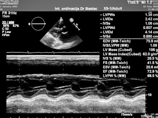
Table 4. 17.07.2026. Tissue Doppler and right
ventricular function -
Weakly expressed but still present sign - ANNULUS REVERSUS.

FOLLOW-UP EXAMINATION 21.07.2026.
Echocardiography still showed preserved left ventricular systolic
function, EF 73% by Teicholz and approximately 60% by Simpson, with
impaired relaxation and reduced compliance. The right ventricle was
LESS dilated, RVSP was 76 mmHg, with TR 3+, while the pericardium
remained thickened, with a thickness of up to 9 mm. Diastolic
dysfunction persists: E/e′ was 21,8. On 21.07.2026, body weight was
68 kg. The patient reports less fatigue and reduced leg edema
[1,13].
Figure 12. 21.07.2026.
At the examination on 21.07.2026 (Figure 12),
preserved left ventricular systolic function was again described,
with a finding interpreted as constrictive pericarditis, with marked
tricuspid regurgitation, pulmonary hypertension and right
ventricular failure.
The combination of clinical systemic congestion, pericardial
thickening and paradoxical septal motion justifies the suspicion of
constrictive physiology, but the morphological change of the
pericardium alone is not sufficient for a definitive diagnosis
[1,2,13].
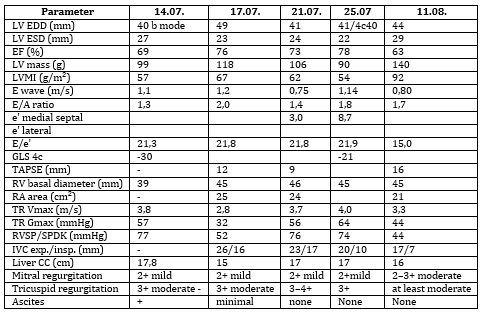
Table 5. Serial echocardiographic follow-up
(14.07–11.08.2026.)
Chest CT scan finding (Dr Stanislav Tadić):
hydrothoraces , pulmonary congestion, no pulmonary infiltrates and
marked hepatomegaly and , splenomegaly, small ascites [1,13].
Serial echocardiographic evaluation initially showed an increase in
pulmonary hypertension with improvement of left ventricular function
and decongestion on 21.07.2026 (RVSP 76 mmHg, TAPSE 9 mm, TR Gmax 56
mmHg), and with excellent clinical improvement (right atrial
dilatation 25 mm and 44 ventricles, significant reduction of
hepatomegaly and of signs of systemic venous congestion..
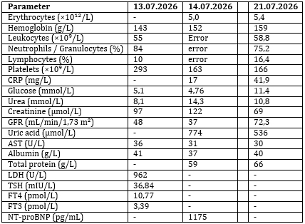
BLOOD LABORATORY ANALYSES
Findings brought from 13.07.2026. - Marked leukocytosis was
recorded. The leukocyte count was 55,0 ×10**9/L, with 84%
granulocytes, while on 21.07.2026. 58,8 ×10**9/L leukocytes were
recorded, with 75,2% granulocytes.
Table 6 . Dynamics of laboratory parameters
(13–21.07.2026.)
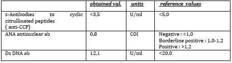
The immunological work-up performed on 23.07.2026
did not show laboratory indicators of a systemic autoimmune disease:
ANA 0,8 COI, anti-CCP <3,5 U/mL and dsDNA 12,1 U/mL.
Table 7. Immunological work-up
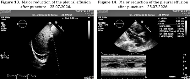
During follow-up until 11.08.2026, a significant
reduction in right ventricular systolic pressure (RVSP or SPDK 44
mmHg) and an improvement in right ventricular function (TAPSE 16 mm)
were recorded, with a reduction in the dimensions of the liver and
the inferior vena cava, i.e. excellent regression of congestion.
Left ventricular diastolic function is improving, E/e' (from 21
to15), and there is a mild ANNULUS REVERSUS SIGN, moderate mitral
regurgitation (2–3+) and residual pulmonary hypertension, indicating
persistent diastolic dysfunction and chronic right heart overload
[1,13].
At the follow-up on 11.08.2026, the clinical condition was
significantly better, with almost complete resolution of peripheral
edema and absence of bilateral pleural effusion on echocardiographic
examination. At the same time, an improvement in right ventricular
function parameters and a reduction in the estimated right
ventricular pressure from 76 to 44 mmHg were recorded.
According to documented measurements, body weight decreased from 77
kg to 52 kg. This weight loss should be interpreted primarily in the
context of marked previous fluid retention and intensive
decongestive therapy. Pleural effusions were no longer recorded
[1,13].
HEMATOLOGIST - However, the hematological problem persisted. On
13.08.2026, a peripheral blood smear showed the presence of all
elements of the granulocytic lineage, with a predominance of
myelocytes and metamyelocytes and blasts up to 2%. Erythrocytes and
platelets were morphologically described as normal, while all
elements of the granulocytic lineage were represented, with a
predominance of myelocytes and metamyelocytes. Blasts accounted for
up to 2%. The finding was assessed as compatible with a
myeloproliferative process, with a recommendation for referral to
the Clinic of Hematology for further diagnostics.
Considering current classifications of myeloid neoplasms, this
finding does not represent a definitive hematological diagnosis, but
rather a basis for further morphological, molecular and clinical
evaluation [17,18].
The finding was assessed as compatible with a myeloproliferative
process, with a recommendation for further diagnostics.
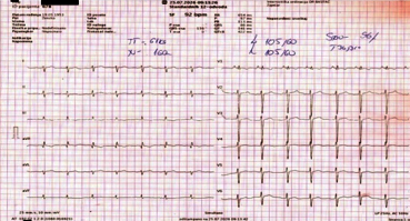
Figure 15. ECG 25 .07. 2026.
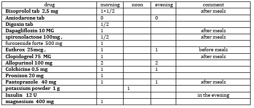
Overview of the oral home therapy given at discharge
from the day hospital of the Dr Bastać Practice (Table 8)
Table 8. oral home therapy 11.08.2026.
Follow-up 03.10.2026. - 80 days after the onset
of the disease
At our invitation, the patient came in, stating that she feels very
well, and denies complaints of significance for the cardiovascular
system. In the period between the last two follow-ups, she was
hospitalized for 2 weeks at the Zaječar General Hospital due to
progression of leukocytosis (CBC during hospitalization Leu 147 x
10*9), but without a final diagnosis and with an indication for
further evaluation by a hematologist, which the patient again
refuses.
Clinical examination - slight deterioration compared to 11 August,
when she was fully cardiac compensated.
Cor: HR 75/min, heart sounds clear, rhythm irregular, frequent SVES,
systolic regurgitant murmur 3/6, p.max over Erb’s point. Pulmo:
breath sounds normal, diminished at the right base, early and late
inspiratory crackles, basal crepitations, Expiration not prolonged.
Abdomen: soft and non-tender on palpation. Liver of soft
consistency, mobile with respiration, palpable 10 cm below the RCM,
spleen of firm consistency, palpable 19 cm below the LCM.
Extremities: doughy edema of the lower legs ++, peripheral arterial
pulses are symmetrically palpable.
CRP 6.4, (normal up to 5 mg/l), mildly elevated
CBC: Hgb106, Er 3.57, Le 65.0, Creatinine 170, GFR 25.6, Uric acid
508, K ion 5.9, HbA1c 5.6, BNP 395. Conclusion: Significant
improvement is maintained, reduced pulmonary hypertension, improved
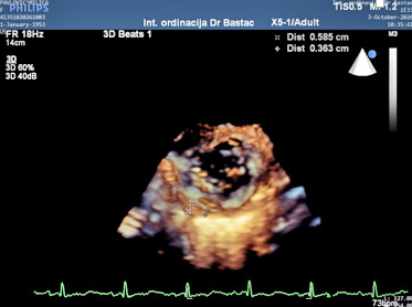
diastolic function, IAS bulging toward the left atrium. TR 3+ is
recorded, the septum makes paradoxical movements. The pericardium is
now of reduced thickness on three-dimensional echocardiography
3D/4D, 3,6 to 5,8 mm, unevenly thickened (also compared to 8 to 9 mm
at the onset of the disease) and separated, without pericardial
effusion.
Mild to moderate mitral regurgitation MR 2+ is recorded. Diastolic
function parameters: impaired relaxation and reduced compliance.
Left ventricle of normal endocavitary dimensions WITHOUT myocardial
hypertrophy and with preserved global systolic function. EF=78%.
Pleural ultrasonography: minimal bilateral pleural effusions. On the
left, pleural effusion in the mid-scapular line basally above the
liver 2cm, in the mid-axillary 5mm, in the posterior axillary 11mm,
in the anterior axillary line 4mm. On the right, effusion in the
mid-scapular line 2,4 cm, posterior axillary line 14 mm, mid-axillary
15 mm
Lung ultrasonography: Basally about 8 B-lines. right and left,
B-lines 7, from the scapular angle A-lines - no congestion.
Since the patient discontinued NSAIDs, corticotherapy and colchicine
on her own initiative, a slight improvement occurred, so it was
explained to the patient that it is very important to take this
therapy for up to 6 months.
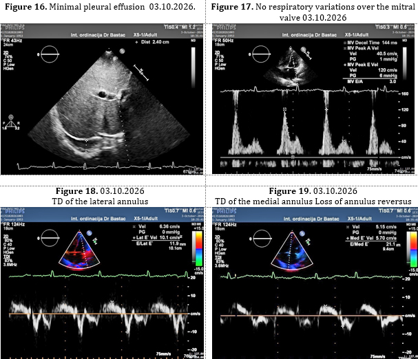
Figure 20. 03.10.2026. Reduction of pericardial
thickness (3D) under anti-inflammatory Th
DISCUSSION
The presented case is characterized by a marked
discrepancy between the preserved left ventricular systolic function
and the severity of the clinical congestion syndrome. During the
most severe phase of the disease, LVEF was preserved, while at the
same time there were left ventricular diastolic dysfunction,
significant tricuspid regurgitation, pulmonary hypertension, right
ventricular dysfunction, right atrial dilatation and signs of
elevated filling pressures.
The key diagnostic problem is distinguishing constrictive
pericarditis from other conditions that can produce a similar
hemodynamic phenotype, primarily HFpEF and restrictive
cardiomyopathy. Constrictive pericarditis can lead to predominant
systemic venous congestion with relatively preserved left
ventricular systolic function, which is why the clinical picture may
be very similar to other forms of right-sided heart failure [1,14].
In this patient, several echocardiographic findings supported the
possibility of constrictive pathophysiology. Particularly notable
were paradoxical movements of the interventricular septum (septal
shift and bounce), pericardial thickening, diastolic filling
abnormalities, dilatation of the right heart chambers and
significant tricuspid regurgitation. At the same time, left
ventricular systolic function remained preserved, with EF values of
approximately 60–76% and normal global longitudinal strain [1,2,13].
The Mayo Clinic echocardiographic criteria include a combination of
respiration-related septal shift, medial e′ and changes in hepatic
venous flow. Combining several criteria increases the specificity
for constrictive physiology. [13]
In the presented case, however, it was not possible to carry out a
complete hemodynamic protocol for definitive confirmation of
constriction. The patient declined further hospitalization, and
invasive pressure assessment was not performed [1,15]. Therefore, it
is more appropriate to designate this finding as
echocardiographically suspected constrictive pericardial
pathophysiology physiology, rather than as definitively confirmed
constrictive pericarditis [1,13].
An important element of the case is the marked leukocytosis. The
leukocyte count was 55,0 ×10⁹/L as early as 13.07.2026, and 58,8
×10⁹/L on 21.07.2026, with marked granulocytosis. In addition,
splenomegaly was present. A later peripheral blood examination
showed the presence of all elements of the granulocytic lineage,
with a predominance of myelocytes and metamyelocytes and blasts up
to 2%, and the finding was designated as compatible with a
myeloproliferative process [17-21].
This finding is significant because it shows that the leukocytosis
was not merely a transient laboratory change during the acute
worsening of heart failure. However, based on the submitted
documentation, it is not possible to definitively determine the type
of hematological disease. Current classifications of myeloid
neoplasms require the integration of clinical, morphological and
molecular data [17,18].
It is precisely in this context that the question arises of a
possible association of the hematological process with the changes
in the pericardium and pleura. In the differential diagnosis,
infiltrative processes, extramedullary hematopoiesis and other
inflammatory or serositis mechanisms may be considered [22-24].
Extramedullary hematopoiesis in the pericardium is a very rare
phenomenon, but it has been described in patients with
myeloproliferative neoplasms. Haroun et al. described extramedullary
hematopoiesis involving the pericardium in a patient with
polycythemia vera, with the development of cardiac tamponade [22].
Mahadevan et al. presented a case of pericardial extramedullary
hematopoiesis with cardiac tamponade and reviewed previously
published cases [23]. In 2025, Hayar et al. described a rare case of
extramedullary hematopoiesis with pericardial involvement in a
patient with a myeloproliferative disease [24].
These data provide a possible pathophysiological framework for
considering a hematological-pericardial association in our case, but
they do not constitute evidence that the pericardial thickening in
the presented patient is a consequence of extramedullary
hematopoiesis. Such a conclusion would require additional
radiological, morphological and/or histological evidence [22-24].
Particularly significant is the therapeutic response to decongestive
therapy, heart failure therapy and anti-inflammatory therapy.
Intensive diuresis and evacuation of the pleural effusions led to a
marked reduction in body weight and resolution of peripheral edema
and pleural effusions. At the same time, a reduction in RVSP from 76
to 44 mmHg and an improvement in TAPSE from 9 to 16 mm were recorded
[1,16]. Such a response confirms the importance of volume overload
in the clinical picture, but by itself neither confirms nor excludes
constrictive pericarditis [1,13]. Also, according to the
recommendations for probable constrictive pericarditis from the
European guideline for myopericardial diseases and the algorithm,
triple anti-inflammatory therapy should be introduced: a
corticosteroid, a nonsteroidal anti-inflammatory drug and colchicine
for up to 6 months. If transient constrictive pericarditis is
established, with clear clinical and imaging improvement and
disappearance of the constrictive pathophysiology, then
pericardiectomy is not required as a definitive treatment measure.
Transient constriction is a form of constrictive pericarditis (CP)
with active inflammation present, which may resolve after
anti-inflammatory therapy or even regress spontaneously. It develops
during pericarditis with or without pericardial effusion
(effusive-constrictive pericarditis). In more than 50% of cases,
constriction resolves after anti-inflammatory therapy [6,7,8]. In
patients with newly diagnosed constrictive pericarditis, a trial of
anti-inflammatory therapy lasting 3 to 6 months is recommended
before referral for pericardiectomy [9,10]. The presence of active
pericardial inflammation was confirmed by inflammatory biomarkers
(C-reactive protein, which was initially 42 mg/L and then fell to 7
mg/L), as well as by cardiovascular magnetic resonance (CMR), which
showed pericardial edema and late gadolinium enhancement (LGE)
[10,11]. In HFpEF with marked congestion and preserved ejection
fraction, the assessment of filling pressures, right ventricular
function and the pulmonary circulation is an important part of the
diagnostic process [16].
For definitive confirmation of constrictive pericarditis in such a
complex presentation, additional modalities would be of importance,
including an ultrafast MDCT scanner or a photon-counting scanner and
cardiovascular magnetic resonance, and, if necessary, right heart
catheterization for direct hemodynamic assessment [1,2,15].
Precisely such further work-up was recommended to the patient, but
it had not been carried out by the last follow-up, because she feels
well and refuses hospitalization at a higher-level institution in
cardiology or hematology.
CONCLUSION and EPILOGUE OF THE CASE:
In patients with marked systemic venous congestion,
hepatosplenomegaly, ascites and anasarca and preserved left
ventricular systolic function, but with signs of severe HFpEF with
reduced left ventricular stroke volume and pulmonary congestion with
pleural effusions, constrictive pericarditis should be considered in
the differential diagnosis [1,13]. Constrictive pericarditis was
suggested by echocardiography: pericardial thickening, parallel
movement of the pericardial layers and paradoxical septal movements
and other Doppler signs of CP [1,2,13]. However, echocardiographic
findings alone are not sufficient for definitive confirmation of the
diagnosis without hemodynamic confirmation. The etiological
diagnosis could be related to the existing chronic
myeloproliferative process [17,18,22-24].
Despite the initial therapeutic failure in this patient with
decompensated heart failure, with huge doses of diuretics, inotropic
support, dobutamine AND corticosteroids, after bilateral pleural
punctures, after 4 days of treatment a gradual recovery occurs. The
peripheral edema and both pleural effusions disappear, as detected
by lung ultrasound. She is later switched to per os th, and there is
a reduction in body weight from 77 kg to 52 kg - a total of 25 kg
over a period of 28 days. A reduction of RVSP to 44 mmHg also
occurs. and an improvement in other echocardiographic dimensions and
parameters [1,16]. At the same time, progressive leukocytosis is
recorded, up to values above 90×10⁹/L, while the examination of the
peripheral blood smear by a hematologist showed a predominance of
myelocytes and metamyelocytes with less than 2% blasts, which was
compatible with a CHRONIC myeloproliferative process. The patient
refuses further treatment at the hematology department of a
higher-level institution and follow-up visits, and in a telephone
conversation claims that she feels well. After almost 2 months,
she comes with moderate deterioration because she discontinued the
anti-inflammatory therapy of her own accord.
References:
-
Schulz-Menger J, Collini V, Gröschel J, Adler Y,
Brucato A, Christian V, et al. 2025 ESC Guidelines for the
management of myocarditis and pericarditis. Eur Heart J.
2025;46(40):3952-4041.
-
Klein AL, Wang TKM, Cremer PC, Abbate A, Adler
Y, Asher C, et al. Pericardial diseases: international position
statement on new concepts and advances in multimodality cardiac
imaging. JACC Cardiovasc Imaging. 2024;17(8):937-88.
-
Klein AL, Abbara S, Agler DA, Appleton CP, Asher
CR, Hoit B, et al. American Society of Echocardiography clinical
recommendations for multimodality cardiovascular imaging of
patients with pericardial disease. J Am Soc Echocardiogr.
2013;26(9):965-1012.
-
Talreja DR, Edwards WD, Danielson GK, Schaff HV,
Tajik AJ, Tazelaar HD, et al. Constrictive pericarditis in 26
patients with histologically normal pericardial thickness.
Circulation. 2003;108(15):1852-7.
-
Adler Y, Charron P, Imazio M, Badano L,
Barón-Esquivias G, Bogaert J, et al. 2015 ESC Guidelines for the
diagnosis and management of pericardial diseases. Eur Heart J.
2015;36(42):2921-64.
-
Imazio M, Brucato A, Maestroni S, Cumetti D,
Belli R, Trinchero R, et al. Risk of constrictive pericarditis
after acute pericarditis. Circulation. 2011;124(11):1270-5.
-
Sagristà-Sauleda J, Permanyer-Miralda G,
Candell-Riera J, Angel J, Soler-Soler J. Transient cardiac
constriction: an unrecognized pattern of evolution in effusive
acute idiopathic pericarditis. Am J Cardiol. 1987;59(9):961-6.
-
Haley JH, Tajik AJ, Danielson GK, Schaff HV,
Mulvagh SL, Oh JK. Transient constrictive pericarditis: causes
and natural history. J Am Coll Cardiol. 2004;43(2):271-5.
-
Sato K, Ayache A, Kumar A, Cremer PC, Griffin B,
Popovic ZB, et al. Improvement in left ventricular mechanics
following medical treatment of constrictive pericarditis. Heart.
2021;107(10):828-35.
-
Feng D, Glockner J, Kim K, Martinez M, Syed IS,
Araoz P, et al. Cardiac magnetic resonance imaging pericardial
late gadolinium enhancement and elevated inflammatory markers
can predict the reversibility of constrictive pericarditis after
antiinflammatory medical therapy: a pilot study. Circulation.
2011;124(17):1830-7.
-
Cosyns B, Plein S, Nihoyannopoulos P, Smiseth O,
Achenbach S, Andrade MJ, et al. European Association of
Cardiovascular Imaging (EACVI) position paper: multimodality
imaging in pericardial disease. Eur Heart J Cardiovasc Imaging.
2015;16(1):12-31.
-
Verhaert D, Gabriel RS, Johnston D, Lytle BW,
Desai MY, Klein AL. The role of multimodality imaging in the
management of pericardial disease. Circ Cardiovasc Imaging.
2010;3(3):333-43.
-
Welch TD, Ling LH, Espinosa RE, Anavekar NS,
Wiste HJ, Lahr BD, et al. Echocardiographic diagnosis of
constrictive pericarditis: Mayo Clinic criteria. Circ Cardiovasc
Imaging. 2014;7(3):526-34.
-
Garcia MJ. Constrictive pericarditis versus
restrictive cardiomyopathy? J Am Coll Cardiol.
2016;67(17):2061-76.
-
Nishimura RA, Carabello BA. Hemodynamics in the
cardiac catheterization laboratory of the 21st century.
Circulation. 2012;125(17):2138-50.
-
McDonagh TA, Metra M, Adamo M, Gardner RS,
Baumbach A, Böhm M, et al. 2023 Focused Update of the 2021 ESC
Guidelines for the diagnosis and treatment of acute and chronic
heart failure. Eur Heart J. 2023;44(37):3627-39.
-
Thiele J, Kvasnicka HM, Orazi A, Gianelli U,
Gangat N, Vannucchi AM, et al. The international consensus
classification of myeloid neoplasms and acute leukemias:
myeloproliferative neoplasms. Am J Hematol. 2023;98(1):166-79.
-
Khoury JD, Solary E, Abla O, Akkari Y, Alaggio
R, Apperley JF, et al. The 5th edition of the World Health
Organization Classification of Haematolymphoid Tumours: Myeloid
and Histiocytic/Dendritic Neoplasms. Leukemia.
2022;36(7):1703-19.
-
Tefferi A, Barbui T. Polycythemia vera and
essential thrombocythemia: 2021 update on diagnosis,
risk-stratification and management. Am J Hematol.
2020;95(12):1599-613.
-
Barbui T, Tefferi A, Vannucchi AM, Passamonti F,
Silver RT, Hoffman R, et al. Philadelphia chromosome-negative
classical myeloproliferative neoplasms: revised management
recommendations from European LeukemiaNet. Leukemia.
2018;32(5):1057-69.
-
Pieri L, Guglielmelli P, Vannucchi AM. The
classification of myeloproliferative neoplasms: current status
and future perspectives. Curr Hematol Malig Rep.
2020;15(4):289-97.
-
Haroun F, Elkis V, Chen A, Lee E. Extramedullary
haematopoiesis presenting with cardiac tamponade in a patient
with polycythaemia vera. BMJ Case Rep.
2017;2017:bcr-2017-221316.
-
Mahadevan NR, Morgan EA, Mitchell RN. Case
report and literature review: cardiac tamponade as a
complication of pericardial extramedullary hematopoiesis.
Cardiovasc Pathol. 2016;25(5):371-4.
-
Hayar SE, Tamir M, Khaldi M, Bouziane M, Haboub
M, Arous S, et al. A rare case of extramedullary hematopoiesis:
when the pericardium produces blood cells. Egypt Heart J.
2025;77(1):86.
|
|
|
|