| |
|
|
INTRODUCTION The human kidneys are small paired organs that
account for only 0.2–0.5% of total body mass, while receiving as
much as 20–25% of the cardiac output. This intensive perfusion,
together with the complex structure and function of the renal
tubules, makes the kidneys particularly susceptible to the harmful
effects of various drugs and chemicals. Most drugs and their
metabolites are eliminated through glomerular filtration and/or
tubular secretion. The elimination process is based on the capacity
for drug metabolism through the oxidative-reductive phase of
biotransformation, as well as on the conjugation of metabolites with
specific carriers that facilitate their excretion into the urine
[1–3].
Nephrotoxicity is defined as a rapid deterioration of kidney
function caused by the toxic effects of substances, most commonly
drugs, which may lead to acute kidney injury (AKI) and necessitate
discontinuation of therapy or even hemodialysis [4]. The proximal
tubules are the nephron segment most susceptible to the nephrotoxic
effects of drugs because of their intense metabolic activity, which,
together with a high osmotic gradient and the presence of transport
mechanisms, facilitates the accumulation of toxins and the
development of injury [2].
Given the increasingly widespread use of drugs in modern therapy,
understanding the mechanisms of nephrotoxicity and recognizing
potentially harmful substances are of crucial importance for the
prevention of kidney injury and the preservation of renal function.
EPIDEMIOLOGY
Drug-induced nephrotoxicity is currently considered to account
for approximately 20% of all cases of nephrotoxicity, with
hospitalized patients and those admitted to intensive care units
being particularly vulnerable. Among older adults, the incidence
reaches up to 66% owing to numerous comorbidities, polypharmacy, and
more frequent exposure to high-risk diagnostic and therapeutic
procedures [5,2].
Nephrotoxicity may manifest as injury affecting different segments
of the nephron and represents one of the etiopathogenetic factors of
acute kidney injury (AKI) and chronic kidney disease (CKD).
According to the results of prospective cohort studies, nephrotoxic
drugs are responsible for AKI in 14–26% of adults, whereas
cross-sectional studies have reported rates of up to 37.5% [2,6].
Approximately 20% of all cases require kidney replacement therapy,
with an associated mortality rate of 60%, even in developed
countries [7].
Risk Factors
Risk factors for drug-induced nephrotoxicity include patient-related
factors, kidney-related factors, and drug-related factors (Table 1).
Table 1. Risk factors for the development of
drug-induced kidney disease [2]
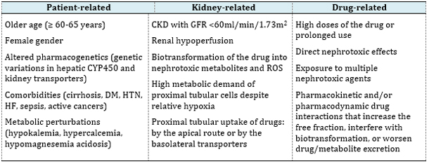
Legend: CYP450, cytochrome P450; DM, diabetes
mellitus; HTN, arterial hypertension; HF, heart failure; CKD,
chronic kidney disease; GFR, glomerular filtration rate;
Patient-related risk factors may be non-modifiable
(e.g., age, sex, and genetic variations) or at least partially
modifiable, as in the case of chronic comorbidities and metabolic
disorders. Both older age and female sex are associated with reduced
lean body mass, lower total body water content, and decreased serum
albumin concentrations, which may contribute to an increased free
fraction of the drug. Furthermore, older individuals have been shown
to have higher circulating levels of angiotensin II and endothelin,
which contribute to vasoconstriction of the efferent arterioles,
increased glomerular filtration, and a greater amount of drugs and
metabolites reaching the renal tubules [3].
An increased risk of nephrotoxicity is undoubtedly associated with
pre-existing CKD with a glomerular filtration rate (GFR) below 60 mL/min,
as well as renal hypoperfusion, whether resulting from true or
effective volume depletion. Additional contributing factors include
intrarenal drug biotransformation, uptake of drugs by proximal
tubular cells, and specific metabolic characteristics of tubular
cells under hypoxic conditions.
One of the most important risk factors is the inherent
nephrotoxicity of certain drugs. Some drugs, such as aminoglycosides,
possess a high nephrotoxic potential and may cause kidney injury
even in patients with minimal or no additional risk factors. In most
cases, drug-induced nephrotoxicity is dose-dependent and/or
associated with prolonged treatment duration. Such reactions are
predictable because they are based on the known pharmacological
properties of the drug and may be attenuated by dose reduction or
drug discontinuation. Less frequently, nephrotoxicity develops as an
unpredictable adverse reaction resulting from an immune-mediated
response that is independent of dose and usually requires complete
withdrawal of the offending drug for resolution of symptoms [3,5,8].
Exposure to multiple nephrotoxic agents and drug interactions
affecting their pharmacokinetic and/or pharmacodynamic properties
further increase the risk of nephrotoxicity.
CURRENT CLASSIFICATIONS OF DRUG-INDUCED
NEPHROTOXICITY
Before 2002, the reported incidence of acute renal
failure varied considerably due to the lack of a standardized
definition. Since then, the RIFLE classification (Risk, Injury,
Failure, Loss, End-stage Kidney Disease) has been developed and was
first published in 2004 [9]. Soon thereafter, it became evident that
even small increases in serum creatinine are clinically significant
because they are associated with higher morbidity and mortality.
Consequently, in 2007, the Acute Kidney Injury Network (AKIN)
modified the RIFLE criteria (AKIN criteria) and introduced the term
AKI instead of acute renal failure [10].
Classification of Drug-Induced AKI According to the Site of Injury
An important initial step in the evaluation of AKI is determining
the primary site of injury. Accordingly, AKI can be classified as
prerenal, renal (intrinsic), or postrenal (Table 2).
• Prerenal injury occurs primarily as a consequence of renal
hypoperfusion. In the context of drug-induced nephrotoxicity,
prerenal AKI may result from the use of nonsteroidal
anti-inflammatory drugs (NSAIDs), calcineurin inhibitors (CNIs), and
modulators of the renin–angiotensin–aldosterone system (RAAS).
• Renal or intrinsic injury may result from damage to any component
of the kidney, including the renal tubules, glomeruli, vascular
structures, interstitium, or tubular lumen. Acute tubular necrosis (ATN)
and acute interstitial nephritis (AIN) are among the most common
forms of intrinsic kidney injury, and anti-infective agents are the
most frequent causative drugs.
• Postrenal injury resulting from obstruction leads to
hydronephrosis and subsequent damage to the renal parenchyma. Drugs
associated with tubular obstruction include acyclovir, methotrexate,
and several antiretroviral agents [11].
Table 2. Potential causes of AKI and recovery
expectations [11]
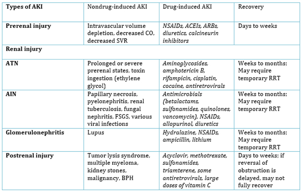
Legend: AKI, acute kidney injury; ATN,
acute tubular necrosis; AIN, acute interstitial nephritis; CO,
cardiac output; SVR, systemic vascular resistance; FSGS, focal
segmental glomerulosclerosis; BPH, benign prostatic hyperplasia;
NSAIDs, non-steroidal anti-inflammatory drugs; ACEIs, angiotensin-converting
enzyme inhibitors; ARBs, angiotensin-II receptor blockers; RRT,
renal replacement therapy);
Classification of Drug-Induced Nephrotoxicity According to
Clinical Presentation (Four Phenotypes)
Despite the previously described classification systems, uniform
standards for the identification of drug-induced nephrotoxicity were
still lacking, and kidney injury often remained unrecognized.
Therefore, in 2015, Mehta et al. proposed four phenotypes of
drug-induced kidney disease according to the underlying mechanism of
injury: acute kidney injury (AKI), glomerular disorder, tubular
dysfunction, and nephrolithiasis.
The clinical presentation of each phenotype is based on changes in
biomarkers and other evidence of kidney injury, including serum
creatinine (AKI), proteinuria or hematuria (glomerular disorders),
electrolyte abnormalities (tubular dysfunction), and
ultrasonographic findings (nephrolithiasis). In order to standardize
the initial phenotypic classification, primary and secondary
criteria were developed, with at least one primary criterion being
required to verify drug-induced nephrotoxicity (Table 3).
Drugs differ in the time course of nephrotoxic manifestations. Some
agents cause acute injury (e.g., aminoglycosides and cephalosporins),
whereas others are associated with a slower and more insidious onset
(e.g., lithium). Taking into account conceptual models of the
temporal evolution of kidney injury, Mehta classified drug-induced
kidney disease into three categories: acute (1–7 days), subacute
(8–90 days), and chronic (>90 days) [12].
Table 3. Primary and secondary criteria for the
four main phenotypes [12]
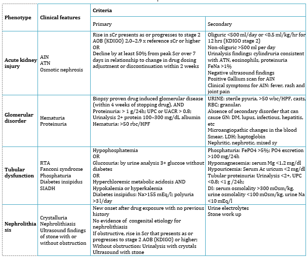
Legend: AIN, acute interstitial nephritis;
ATN, acute tubular necrosis; DM, diabetes mellitus; FeNa, fractional
excretion of sodium; FePO4, fractional phosphorus excretion, GN,
glomerulonephritis; HPF, high powered field; LDH, lactate
dehydrogenase; SIADH, syndrome of inadequate antidiuretic hormone
secretion; UPC, urinary protein/creatinine ratio; UACR, urinary
albumin/creatinine ratio;
Biomarker-Based Classification of Nephrotoxicity
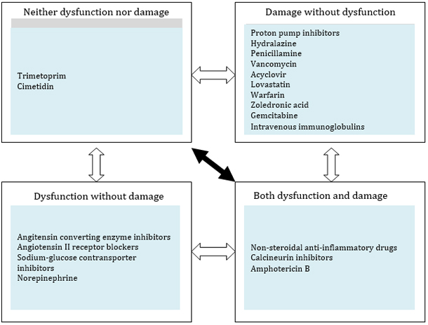
The following conceptual classification was proposed in 2019 by
an international expert panel composed of nephrologists and
pharmacists. This classification integrates both functional
biomarkers and biomarkers of kidney damage, together with the
predominant mechanisms of nephrotoxicity, and categorizes drugs into
four groups: dysfunction without damage, damage without dysfunction,
both dysfunction and damage, and neither dysfunction nor damage.
Examples of drugs belonging to each category are provided to further
illustrate this classification system, while possible transitions
between categories are shown by arrows (Figure 1).
- Neither dysfunction nor damage
This category includes drugs that cause an increase in serum
creatinine without inducing kidney dysfunction or structural
damage. For example, some drugs may reduce tubular creatinine
secretion or increase creatinine production without affecting
kidney function. These drugs are not truly nephrotoxic, and this
clinical situation is referred to as pseudo-AKI.
- Dysfunction without damage
This category includes drugs that impair kidney function by
affecting systemic or intraglomerular hemodynamics. By reducing
renal perfusion, they decrease intraglomerular pressure,
filtration fraction, and GFR, resulting in an increase in serum
creatinine concentration. In some cases, this alteration in
glomerular hemodynamics represents an adverse effect, whereas in
others it reflects the intended therapeutic action of the drug.
The most common examples are RAAS modulators and vasoactive
agents.
- Damage without dysfunction
Kidney damage within this category may involve the glomeruli,
tubules, or interstitium. Drugs may induce direct or indirect
tubular damage/injury through crystal and cast formation,
oxidative stress, inflammation, mitochondrial dysfunction, and
immune-mediated reactions.
- Both dysfunction and damage
This category includes drugs associated with both hemodynamic
and non-hemodynamic mechanisms of AKI development.
Non-hemodynamic features of AKI include glomerular, tubular, and
interstitial injury occurring either alone or in combination. A
given drug may induce functional changes through one mechanism
and tissue damage through another, or both may result from the
same pathogenic pathway. Typical examples are NSAIDs and CNIs
[13].
Algorithm 1. Four categories of nephrotoxicity
based on functional and damage biomarkers [13]
Biomarkeri disfunkcije i oštećenja
Biomarkers of Dysfunction and Injury
Drug-induced nephrotoxicity, similarly to other kidney diseases, can
be diagnosed using simple blood and/or urine tests, including the
measurement of serum creatinine and blood urea levels, as well as
assessment of GFR using creatinine clearance. However, these tests
are known to be imperfect markers of kidney function, as they are
influenced by numerous renal and extrarenal factors.
Owing to continuous research efforts, novel biomarkers have been
identified that are more sensitive and capable of detecting kidney
injury at an earlier stage. Functional biomarkers include cystatin
C, proenkephalin A (PENK), beta-trace protein (BTP), and neutrophil
gelatinase-associated lipocalin (NGAL), which are measured in
plasma. In addition, urinary biomarkers have been identified that
may indicate injury to specific nephron segments. However, further
studies are required before their routine clinical application can
be recommended. Such studies should address several issues of
crucial importance: when these biomarkers should be measured,
whether they should be used to guide clinical decision-making, and
what actions should be taken when their levels are elevated [14].
DRUG-INDUCED KIDNEY DISEASES
Acute Tubular Necrosis
ATN is a common cause of AKI in critically ill patients and is most
often a consequence of prolonged prerenal insults or exposure to
nephrotoxins. Histological changes demonstrate necrosis of the renal
tubular epithelium, with necrotic cells being shed into the tubular
lumen, where they may cause extensive tubular injury. Regeneration
is possible if the tubular basement membrane remains intact. The
injury is usually reversible, although it may persist for days or
weeks, and temporary renal replacement therapy (RRT) may be
required. If ischemia is severe and prolonged, the injury may become
irreversible [15,11].
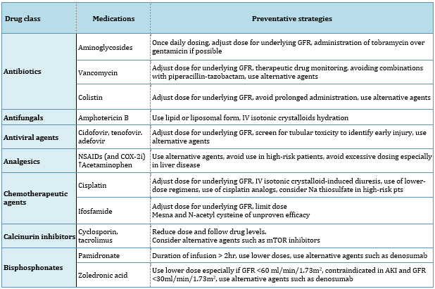
Direct tubular injury may be caused by various classes of drugs,
including antimicrobial agents, chemotherapeutic drugs, calcineurin
inhibitors, bisphosphonates, and contrast media. Preventive
strategies for many of these agents include volume expansion to
correct hypovolemia and increase tubular flow, adjustment of drug
dosage according to baseline GFR, and the use of alternative agents
(e.g., mTOR inhibitors instead of calcineurin inhibitors or
denosumab instead of bisphosphonates). For certain drugs, such as
vancomycin and CNIs, therapeutic drug monitoring is required.
Specific measures for vancomycin include avoidance of concomitant
administration with piperacillin–tazobactam, whereas the
nephrotoxicity of amphotericin B is significantly reduced when
lipid-based or liposomal formulations are used (Table 4) [6].
Table 4. Drugs associated with acute tubular
injury and preventative strategies [6]
Legend: IV, intravenous; NSAIDs nonsteroidal
anti-inflammatory drugs; COX, cyclo-oxygenase; mTOR, mammalian
target of rapamycin;
Acute Interstitial Nephritis
AIN is an immune-mediated form of kidney injury, with drugs being
responsible for more than 70% of cases (drug-induced acute
interstitial nephritis, DI-AIN) [17]. Kidney biopsies performed in
the evaluation of AKI indicate that the incidence of DI-AIN is
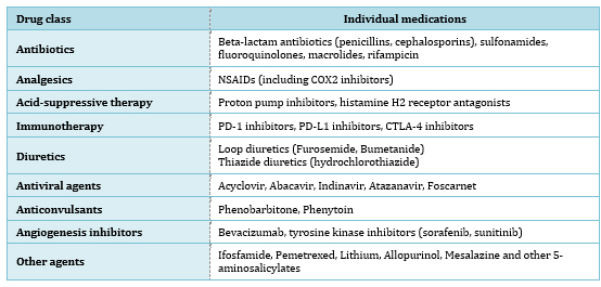
approximately 15%. To date, more than 250 drugs have been reported
to cause AIN, with antibiotics, NSAIDs, proton pump inhibitors (PPIs),
and immune checkpoint inhibitors being the most commonly implicated
agents (Table 5) [6,17,18].
Table 5. Drugs associated with acute interstitial
nephritis
Legend: NSAIDs nonsteroidal
anti-inflammatory drugs; COX, cyclo-oxygenase; PD-1i, Programmed
Death-1 inhibitors; PD-L1i, Programmed Death Ligand-1 inhibitors;
CTLA-4i, Cytotoxic T-Lymphocyte Antigen-4 inhibitors;
DI-AIN presents either as AKI or as a subacute decline in kidney
function, indicating that symptoms may develop within a few days or
several weeks to months after exposure to the drug that triggered
the inflammatory cascade. The classic hypersensitivity triad
consisting of rash, fever, and eosinophilia is observed only rarely
(<10%). More commonly, patients present with nonspecific symptoms
such as fatigue, malaise, and nausea, occasionally accompanied by
flank pain. Many patients develop hypertension due to increased
angiotensin II activity and the resulting arterial vasoconstriction
in the setting of interstitial inflammation. Patients are usually
non-oliguric unless advanced AKI develops, and some may even be
polyuric because of impaired urinary concentrating ability [18,19].
Nephrotic syndrome may occur in cases induced by NSAIDs, although
proteinuria of up to 1 g/24 h, sterile pyuria with leukocyte casts,
and microscopic hematuria are more common findings. Although
laboratory findings may support a high clinical suspicion of DI-AIN,
these tests have limited sensitivity and specificity; therefore, the
definitive diagnosis is established by kidney biopsy. Novel urinary
biomarkers may provide a noninvasive diagnostic alternative in the
future.
Histopathological findings indicate involvement of both the renal
tubules and the interstitium. The characteristic feature is an
extensive interstitial inflammatory infiltrate composed
predominantly of lymphocytes and monocytes, although eosinophils,
plasma cells, histiocytes, and polymorphonuclear leukocytes may also
be present, and less commonly granulomas may be observed. Initially,
the inflammatory infiltrate may be associated with interstitial
edema; however, over time, interstitial fibrosis and tubular atrophy
develop, findings that are characteristic of chronic interstitial
nephritis [19,20].
Treatment and Prognosis of Drug-Induced Acute Interstitial Nephritis
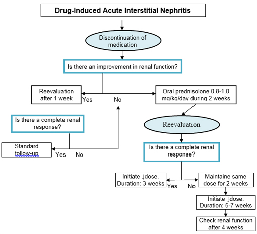
Discontinuation of the suspected drug is generally considered the
cornerstone of DI-AIN treatment, although identification of the
offending agent may sometimes be challenging. Reassessment of serum
creatinine is recommended one week after withdrawal of the suspected
drug. In the absence of significant improvement, treatment with oral
prednisolone at a dose of 0.8–1 mg/kg/day is recommended for 2
weeks. If a complete renal response is achieved, tapering of
corticosteroid therapy should be initiated. In patients with an
incomplete response, the same dose of prednisolone is recommended
for an additional 2 weeks, followed by gradual dose reduction over a
period of 5–7 weeks (Algorithm 1) [20].
Early reports of methicillin-associated AIN suggested that the
disease was relatively benign and associated with a favorable
prognosis. However, this has proven to be far from reality, as
incomplete recovery of kidney function is observed in approximately
40% of patients [21], and up to one-third of patients require
dialysis treatment. Older adults are not only at a higher risk of
developing the disease but also tend to have a poorer prognosis
[20]. Prolonged impairment of kidney function is considered an
unfavorable prognostic factor, whereas early diagnosis and timely
initiation of treatment are associated with more favorable outcomes
[22]. Histopathological findings undoubtedly have predictive value
despite the inconsistencies reported in clinical studies: diffuse
cellular infiltration, a greater degree of interstitial fibrosis and
tubular atrophy, and the presence of granulomas have all been
associated with a less favorable prognosis) [20].
Algorithm 1. Algorithm for the treatment of acute
interstitial nephritis caused by drugs [20]
Crystal Nephropathies Caused by Drugs
Crystal nephropathies are an important but often underrecognized
cause of kidney disease and are characterized histologically by the
presence of intrarenal crystal deposits that primarily affect the
tubulointerstitial compartment. Clinically, patients present with
urinary abnormalities such as crystalluria, proteinuria, and urinary
casts, although tubulopathies, AKI, and CKD may also develop
[23,24,25]. Microscopic identification of various pathological
crystals and/or crystal-containing casts in the urine suggests the
possibility of crystal nephropathy. Demonstration of crystals within
the kidney by biopsy is crucial for establishing the diagnosis and
requires evaluation of the underlying cause.
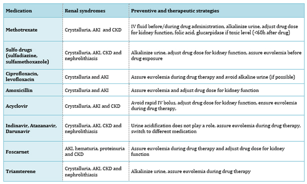
Drug-induced intrarenal crystal deposition occurs primarily as a
consequence of renal excretion of the drug or its metabolites and
drug supersaturation in urine. Conditions that favor crystal
formation include hypovolemia/dehydration (resulting in reduced
urinary flow), drug overdose (leading to increased urinary drug
concentrations), and urinary pH, the effect of which depends on the
pKa of the respective drug (acidic urine for methotrexate and
sulfadiazine, and alkaline urine for indinavir, atazanavir, and
ciprofloxacin). The pathophysiological mechanisms involved in the
development of drug-induced crystal nephropathy include substrate
supersaturation, nucleation, aggregation, further crystal growth,
cellular membrane injury, intratubular obstruction, direct and
indirect cytotoxicity, inflammation, and necroinflammation [25]. The
drugs most commonly associated with these processes are listed in
Table 6, together with their clinical presentation and the basic
preventive and therapeutic strategies.
Table 6. Drug-induced crystalline nephropathies
[25]
Legend: AKI, acute kidney injury; CKD,
chronic kidney disease; IV, intravenous;
Drug-Induced Rhabdomyolysis
Rhabdomyolysis is a clinical syndrome in which skeletal muscle
injury leads to myocyte lysis and the release of intracellular
contents into the systemic circulation, including electrolytes,
purines, enzymes such as creatine kinase (CK), and myoglobin [26].
Myoglobin is considered responsible for renal toxicity through three
principal mechanisms: renal vasoconstriction, formation of
intratubular casts, and direct toxicity to renal tubular cells [27].
The typical signs and symptoms of rhabdomyolysis are known as the
classic triad and include acute or subacute myalgia, muscle weakness
predominantly affecting the proximal muscles of the lower
extremities, and dark tea-colored urine. Clinical manifestations
range from subclinical elevations of serum CK, lactate dehydrogenase
(LDH), and aspartate aminotransferase (AST) to severe electrolyte
disturbances, cardiac arrhythmias, AKI, and disseminated
intravascular coagulation [28].
The most common causes of rhabdomyolysis in adults include strenuous
physical activity, illicit drugs (heroin, cocaine, methadone, and
methamphetamine), alcohol abuse, medications, muscle diseases,
trauma, neuroleptic malignant syndrome, seizures, and prolonged
immobilization [29]. Drugs most commonly implicated in the
development of rhabdomyolysis include statins, fibrates,
sedative-hypnotics, anesthetic agents associated with malignant
hyperthermia, neuroleptics, anabolic steroids/corticosteroids, and
newer-generation anticonvulsants [26,28]. Treatment consists of
discontinuation of the offending drug and restoration of
intravascular volume in order to achieve a urine output exceeding 2
L/24 h. At present, there is insufficient evidence to support the
routine use of loop diuretics, mannitol, or bicarbonate in the
treatment of rhabdomyolysis. In patients with severe AKI and other
life-threatening complications, such as hyperkalemia, hypocalcemia,
or volume overload, RRT should be considered [30].
Drug-Induced Thrombotic Microangiopathy
Drug-induced thrombotic microangiopathy (TMA) accounts for 10–13% of
all cases of TMA and approximately 20–30% of secondary forms, which
are considerably more common than primary forms (80–90%). The
classic triad of microangiopathic hemolytic anemia,
thrombocytopenia, and kidney injury is rarely observed in its
entirety. More frequently, patients present with subacute forms
characterized by mild thrombocytopenia and a gradual decline in
kidney function, or even with kidney-limited TMA, which is
associated with a more favorable prognosis [31]. The key
pathological feature is endothelial injury caused by heterogeneous
mechanisms, most commonly involving an idiosyncratic acute
antibody-mediated immune response or a direct toxic effect of the
drug related to dosage or duration of therapy [32,33]. The list of
drugs potentially associated with TMA is rapidly expanding and
includes numerous antimicrobial agents, immunosuppressive drugs,
chemotherapeutic agents, antiplatelet drugs, as well as various
other medications and illicit substances [31,32,33].
The cornerstone of treatment is discontinuation of the suspected
drug and supportive care. Plasma exchange and rituximab should be
considered only in patients with suspected immune-mediated TMA,
particularly in cases potentially associated with ticlopidine or in
patients who fail to improve after withdrawal of the offending drug.
In severe cases, complement blockade with the monoclonal antibody
eculizumab may be used as rescue therapy. By inhibiting complement
factor C5, eculizumab prevents the formation of the membrane attack
complex and thereby inhibits cell lysis and death [31].
(POTENTIALLY) NEPHROTOXIC DRUGS IN CLINICAL PRACTICE
Aminoglycosides
The nephrotoxicity of aminoglycosides is well recognized (occurring
in 10–25% of cases); however, these agents continue to be used in
the treatment of severe Gram-negative infections and infective
endocarditis. In recent years, their use has even increased because
of the progressive resistance to other antimicrobial agents and the
lack of new therapeutic alternatives [34]. The mechanisms underlying
aminoglycoside-induced nephrotoxicity include interaction with
proximal tubular cells leading to cellular apoptosis and necrosis,
as well as a reduction in renal blood flow resulting in parenchymal
ischemia and promoting the development of AKI. Nephrotoxicity most
commonly manifests as non-oliguric AKI, usually occurring 5–7 days
after the initial exposure to the drug, or as proximal tubular
dysfunction known as Fanconi syndrome, which is characterized by
metabolic acidosis, hypophosphatemia, glycosuria, and aminoaciduria
[6,35].
Strategies for the safer use of aminoglycosides have been defined in
clinical guidelines, which recommend that:
- aminoglycosides should not be used for the treatment of
infections unless no suitable alternative is available;
- in otherwise healthy and clinically stable patients,
aminoglycosides should be administered as a single daily dose
rather than in multiple daily doses;
- aminoglycoside serum concentrations should be monitored when
multiple daily dosing is continued for more than 24 hours or
when once-daily dosing is continued for more than 48 hours;
- topical or local administration of aminoglycosides (e.g.,
respiratory aerosols or instilled antibiotic beads) should be
preferred over intravenous administration whenever feasible and
clinically appropriate [36].
Treatment of aminoglycoside-induced nephrotoxicity consists of
supportive care and discontinuation of the offending drug. Following
drug withdrawal, the prognosis is generally favorable, and recovery
of kidney function usually occurs within several days to several
weeks. However, pre-existing kidney disease, advanced age, and
prolonged exposure to aminoglycosides are associated with a high
risk of incomplete recovery and progression to CKD. On the other
hand, aminoglycoside-induced AKI in critically ill patients is
associated with increased morbidity and mortality, particularly when
complicated by sepsis, hypotension, or multiple organ dysfunction
[37].
Nonsteroidal Anti-inflammatory Drugs
NSAIDs exert their analgesic, anti-inflammatory, and antipyretic
effects through inhibition of cyclooxygenase (COX), the enzyme
responsible for converting arachidonic acid released from cell
membranes into various eicosanoids, including thromboxane,
prostaglandin E2, and prostacyclin. Within the kidney,
prostaglandins exert vasodilatory effects and influence the tubular
transport of water and electrolytes by reducing sodium and water
reabsorption and increasing potassium secretion [38]. Prostaglandin
production is increased in CKD as a mechanism aimed at improving
perfusion of the remaining nephrons (“prostaglandin-dependent
state”). Therefore, in these patients, both selective and
non-selective COX inhibition are considered equally hazardous.
Current clinical guidelines recommend avoiding prolonged NSAID use
when the glomerular filtration rate is 30–60 mL/min/1.73 m² and
complete avoidance when the glomerular filtration rate is below 30
mL/min/1.73 m² [39].
The nephrotoxic effects of NSAIDs may manifest clinically in several
ways:
• AKI, predominantly mediated by hemodynamic mechanisms, leading
either to a reversible decline in GFR or to ischemic tubular injury;
• Fluid and electrolyte disorders, including hyperkalemia,
occasionally metabolic acidosis, hyponatremia, and sodium and water
retention, resulting in edema, worsening of congestive heart
failure, and hypertension;
• AIN (NSAIDs account for 10–15% of cases);
• Proteinuria and nephrotic syndrome resulting from membranous
nephropathy or minimal change disease as less common complications;
• Risk of CKD progression associated with cumulative drug exposure
[38].
The use of NSAIDs is generally not considered excessively harmful in
young individuals without kidney disease or significant
comorbidities. However, because their nephrotoxic effects are
dose-dependent, particular attention should be paid to chronic use,
which increases the risk of nephrotoxicity. In CKD, NSAIDs directly
affect kidney function through inhibition of prostaglandin
synthesis; therefore, the indications for their use should be
carefully evaluated, taking into account existing risk factors and
the expected clinical benefit [40]. Risk factors for NSAID-induced
nephrotoxicity are presented in Table 7 [38].
Table 7. Risk factors for NSAID-induced
nephrotoxicity [38]:
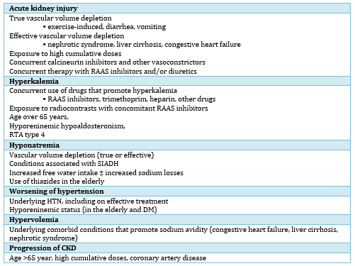
Legend: RAAS, renin-angiotensin-aldosterone
system; RTA, renal tubular acidosis; SIADH, syndrome of inadequate
antidiuretic hormone secretion; HTN, arterial hypertension; DM,
diabetes mellitus;
Renin–Angiotensin–Aldosterone System Inhibitors
The nephroprotective effects of RAAS inhibitors are well
established, particularly in patients with kidney disease and
diabetes mellitus. These effects are mediated through blood pressure
reduction, slowing of GFR decline, reduction of proteinuria, and
delay of progression to advanced stages of CKD [2,41]. Initial
decline in eGFR after RAAS inhibitor initiation should not
automatically be interpreted as nephrotoxicity. These drugs are
rarely truly nephrotoxic, but their effects on intrarenal
hemodynamics may result in a reversible decline in GFR and
consequently AKI [2].
Such adverse renal effects may occur in patients with pre-existing
impairment of glomerular autoregulation, which depends on
prostaglandin-mediated vasodilation of the afferent arteriole and
angiotensin II-mediated vasoconstriction of the efferent arteriole
[42]. Nephrotoxicity may result from hypoperfusion associated with
low mean arterial pressure (reduced cardiac output or low systemic
vascular resistance), volume depletion (e.g., due to diuretic
therapy), or pre-existing renovascular disease, such as bilateral
renal artery stenosis or severe intrarenal arteriosclerosis [43].
The nephrotoxic potential of RAAS inhibitors may also become
apparent as a result of drug interactions. A typical example is the
concomitant chronic use of NSAIDs and RAAS inhibitors, particularly
in older adults, with an additional increase in risk when a diuretic
is also prescribed. This phenomenon is commonly referred to as the
“triple whammy” interaction [44].
Management of patients with hemodynamic instability and AKI requires
correction of hypovolemia and hypotension; therefore, temporary
discontinuation of all antihypertensive agents, including RAAS
inhibitors, is a logical approach. However, after restoration of an
adequate circulating volume, normalization of blood pressure, and
recovery of GFR, these agents should be reintroduced according to
the original indication for their use [41]. In patients with CKD,
treatment with RAAS inhibitors should be continued unless serum
creatinine increases by more than 30% within four weeks after
initiation of therapy or dose escalation. Dose reduction or
discontinuation of RAAS inhibitors should be considered in the
presence of symptomatic hypotension or uncontrolled hyperkalemia
despite medical treatment, or in order to reduce uremic symptoms
during the management of kidney failure (estimated GFR <15 mL/min/1.73
m²) [45].
Statins
Statins (HMG-CoA reductase inhibitors) play an undisputed role in
the primary and secondary prevention of cardiovascular disease,
including among patients with predialysis CKD [45]. However, they
may also exert nephrotoxic effects, making careful monitoring of
kidney function during the first months of treatment advisable.
Statin-induced nephrotoxicity has been directly associated with high
drug doses and indirectly with interactions involving other agents
that may increase serum statin concentrations [46]. Potential
mechanisms of toxicity include alterations in cell membrane
permeability, reduced ubiquinone levels, and depletion of
isoprenoids resulting from inhibition of cholesterol synthesis [46].
Identified risk factors include advanced age, female sex, liver
dysfunction, and diabetes mellitus. The clinical presentation of
statin-induced nephrotoxicity includes rhabdomyolysis, ranging from
mild elevations in serum creatine kinase levels and myalgia to
severe muscle weakness, muscle cramps, myositis, and rhabdomyolysis.
In addition, statins may cause acute or subacute interstitial
nephritis through immune-mediated mechanisms, which generally
resolves following drug withdrawal and corticosteroid therapy [47].
Recent epidemiological studies have indicated a significantly
increased risk of AKI, particularly among patients initiating
treatment with high-potency statins [48]. However, these cases are
often underreported, which may be explained by the characteristics
of the population receiving these drugs. Patients treated with
statins are generally older and frequently have comorbidities such
as diabetes mellitus, hypertension, and atherosclerotic renovascular
disease. In addition, they often receive concomitant medications,
including RAAS inhibitors and diuretics, all of which may contribute
to renal dysfunction. Furthermore, many patients are treated with
antiplatelet agents, making kidney biopsy more difficult in the
setting of AKI. Finally, statin-induced acute or subacute
tubulointerstitial nephritis may develop insidiously and remain
subclinical, thereby going unrecognized if kidney function is not
routinely monitored [47,49].
Proton Pump Inhibitors
Proton pump inhibitors (PPIs) are among the most commonly used drugs
worldwide, with an estimated 53–69% of prescriptions lacking an
appropriate medical indication [50]. Although PPIs were considered
safe for many years, evidence from observational studies suggests an
association between PPI use and a variety of adverse effects,
particularly adverse renal outcomes, including AKI, AIN, and CKD
[50,51].
AIN may develop within hours to several months after initiation of
therapy and rarely presents with the classic triad of fever, rash,
and eosinophilia. However, sterile pyuria, hematuria, and
eosinophiluria may be detected on urinalysis. A rapid decline in
kidney function may precipitate AKI, or AIN may be complicated by
the development of interstitial fibrosis and tubular atrophy,
resulting in chronic interstitial nephritis that gradually
progresses to CKD. According to available studies, more than half of
patients fail to achieve complete recovery of kidney function
following AIN. Hypomagnesemia has also been reported as an adverse
effect. Although the underlying mechanisms remain insufficiently
understood, available evidence suggests an association with CKD
[50,52].
Preventive measures include limiting excessive PPI use to
appropriate medical indications, with careful attention to both dose
and duration of therapy. Particular caution is warranted in patients
with CKD or those at increased risk of developing CKD, and
alternative therapeutic options should be considered whenever
appropriate. When treatment with PPIs is clearly indicated,
monitoring of GFR and serum magnesium levels is recommended,
especially in patients receiving concomitant diuretic therapy [50].
CONCLUSION
Drug-induced nephrotoxicity is a significant cause
of both acute and chronic kidney injury and represents an important
contributor to morbidity and mortality in contemporary clinical
practice. The risk of kidney injury is particularly pronounced in
older adults, patients with pre-existing CKD, multiple comorbidities,
and polypharmacy. Timely identification of risk factors, rational
use of potentially nephrotoxic drugs, individualized dosing, and
regular monitoring of kidney function constitute the cornerstone of
prevention. Advances in the understanding of pathophysiological
mechanisms and the development of novel biomarkers provide
opportunities for earlier detection of kidney injury and more
precise patient monitoring. Given that a substantial proportion of
drug-induced kidney injuries are potentially preventable, physician
education aimed at the early recognition of nephrotoxicity remains a
key element in preserving kidney function and improving treatment
outcomes.
LITERATURE:
- Kalucki SA, Lardi C, Garessus J, Kfoury A, Grabherr S,
Burnier M, et al. Reference values and sex differences in
absolute and relative kidney size. A Swiss autopsy study. BMC
Nephrol. 2020; 21: 289.
- Dobrek L. A Synopsis of Current Theories on Drug-Induced
Nephrotoxicity. Life (Basel). 2023; 13(2): 325.
- Perazella MA. Pharmacology behind common drug
nephrotoxicities. Clin J Am Soc Nephrol. 2018; 13: 1897–1908.
- Al-Naimi MS, Rasheed HA, Hussien NR, Al-Kuraishy HM, Al-Gareeb
AI. Nephrotoxicity: Role and significance of renal biomarkers in
the early detection of acute renal injury. J Adv Pharm Technol
Res. 2019; 10(3): 95-99.
- Naughton CA. Drug-induced nephrotoxicity. Am Fam Physician.
2008;78(6):743–750.
- Perazella MA, Rosner MH. Drug-induced acute kidney injury.
Clin J Am Soc Nephrol. 2022; 17(8): 1220–1233.
- Kidney Disease: Improving Global Outcomes (KDIGO) Acute
Kidney Injury Work Group. KDIGO clinical practice guideline for
acute kidney injury. Kidney Int Suppl. 2012; 2: 1–138.
- Siddama A., Suneel I.M. Drug induced kidney disease. Open
Acc. J. Toxicol. 2017; 2(5): 555576.
- Bellomo R, Ronco C, Kellum JA, et al. Acute Dialysis Quality
Initiative Workgroup Acute renal failure – definition, outcome
measures, animal models, fluid therapy and information
technology needs: the Second International Consensus Conference
of the Acute Dialysis Quality Initiative (ADQI) Group. Crit
Care. 2004; 8(4): R204–R212.
- Coca SG, Peixoto AJ, Garg AX, Krumholz HM, Parikh CR. The
prognostic importance of a small acute decrement in kidney
function in hospitalized patients: a systematic review and
meta-analysis. Am J Kidney Dis. 2007; 50(5): 712–720.
- McDaniel BL, Bentley ML. The role of medications and their
management in acute kidney injury. Integr Pharm Res Pract. 2015;
4: 21-29.
- Mehta RL, Awdishu L, Davenport A, Murray PT, Macedo E, Cerda
J, et al. Phenotype standardization for drug-induced kidney
disease. Kidney Int. 2015; 88(2): 226-234.
- Karimzadeh I, Barreto EF, Kellum JA, Awdishu L, Murray PT,
Ostermann M, et al. Moving toward a contemporary classification
of drug-induced kidney disease. Crit Care. 2023; 27(1): 435.
- Parikh CR, Koyner JL. Biomarkers in acute and chronic kidney
diseases. In: Chertow G, Luyckx V, Marsden P, Skorecki K, Taal
M, Alan Y, editors. Brenner and Rector’s The Kidney. 11.
Elsevier; 2020.
- Moore EM, Bellomo R, Nichol AD. The meaning of acute kidney
injury and its relevance to intensive care and anaesthesia.
Anaesth Intensive Care. 2012; 40(6): 929–948.
- Perazella MA, Markowitz GS. Drug-induced acute interstitial
nephritis. Nat Rev Nephrol. 2010; 6(8): 461-470.
- Raghavan R, Shawar S. Mechanisms of Drug-Induced
Interstitial Nephritis. Adv Chronic Kidney Dis. 2017; 24(2):
64-71.
- Kodner CM, Kudrimoti A. Diagnosis and management of acute
interstitial nephritis. Am Fam Physician. 2003;67(12):2527-2534.
- Finnigan NA, Rout P, Leslie SW, et al. Allergic and
Drug-Induced Interstitial Nephritis. [Updated 2023 Sep 12]. In:
StatPearls [Internet]. Treasure Island (FL): StatPearls
Publishing; 2026 Jan.
- Sanchez-Alamo B, Cases-Corona C, Fernandez-Juarez G. Facing
the Challenge of Drug-Induced cute Interstitial Nephritis.
Nephron. 2023;147(2):78-90.
- Rossert J, Fischer E. Acute interstitial nephritis.
Comprehensive clinical nephrology. Philadelphia: Elsevier
Limited; 2018.
- Muriithi AK, Leung N, Valeri AM, Cornell LD, Sethi S, Fidler
ME, Nasr SH. Biopsy-proven acute interstitial nephritis,
1993-2011: a case series. Am J Kidney Dis. 2014;64(4):558-566.
- Yarlagadda SG, Perazella MA. Drug-induced crystal
nephropathy: an update. Expert Opin Drug Saf. 2008; 7(2):
147-158.
- Daudon M, Frochot V, Bazin D, Jungers P. Drug-Induced Kidney
Stones and Crystalline Nephropathy: Pathophysiology, Prevention
and Treatment. Drugs. 2018;78(2):163-201.
- Perazella MA, Herlitz LC. The Crystalline Nephropathies.
Kidney Int Rep. 2021;6(12):2942-2957.
- Keltz E, Khan FY, Mann G. Rhabdomyolysis. The role of
diagnostic and prognostic factors. Muscles Ligaments Tendons J.
2014;3(4):303-312.
- Petejova N, Martinek A. Acute kidney injury due to
rhabdomyolysis and renal replacement therapy: a critical review.
Crit Care. 2014;18(3):224.
- Deng Z, Wang S, Wu C. Rhabdomyolysis associated with
newer-generation anti-seizure medications (ASMs): a real-world
retrospective and pharmacovigilance study. Front Pharmacol.
2023; 4:1197470.
- Melli G, Chaudhry V, Cornblath DR. Rhabdomyolysis: an
evaluation of 475 hospitalized patients. Medicine (Baltimore).
2005;84(6):377-385.
- Yang BF, Li D, Liu CL, Luo Y, Shi J, Guo XQ, et al. Advances
in rhabdomyolysis: A review of pathogenesis, diagnosis, and
treatment. Chin J Traumatol. 2026;29(1):21-31.
- Mazzierli T, Allegretta F, Maffini E, Allinovi M.
Drug-induced thrombotic microangiopathy: An updated review of
causative drugs, pathophysiology, and management. Front
Pharmacol. 2023;13:1088031.
- Al-Nouri ZL, Reese JA, Terrell DR, Vesely SK, George JN.
Drug-induced thrombotic microangiopathy: a systematic review of
published reports. Blood. 2015;125(4):616-618.
- Abou-Ismail MY, Kapoor S, Citla Sridhar D, Nayak L, Ahuja S.
Thrombotic microangiopathies: An illustrated review. Res Pract
Thromb Haemost. 2022;6(3):e12708.
- Lopez-Novoa JM, Quiros Y, Vicente L, Morales AI,
Lopez-Hernandez FJ. New insights into the mechanism of
aminoglycoside nephrotoxicity: an integrative point of view.
Kidney Int. 2011; 79(1): 33-45.
- Campbell RE, Chen CH, Edelstein CL. Overview of
Antibiotic-Induced Nephrotoxicity. Kidney Int Rep.
2023;8(11):2211-2225.
- Khwaja A. KDIGO clinical practice guidelines for acute
kidney injury. Nephron Clin Pract. 2012;120(4):c179-184.
- Chowdary PJ, Kumar AS, Kumari NH, Sowjanya KK, Sravya D,
Divakar D. A comprehensive review on drug-induced (aminoglycosides)
acute kidney injury: mechanism, risk factors, diagnosis, and
therapeutic approaches. World J. Adv. Pharm. Sci.
2025;2(2):73-80.
- Baker M, Perazella MA. NSAIDs in CKD: Are They Safe? Am J
Kidney Dis. 2020;76(4):546-557.
- Shlipak MG, Tummalapalli SL, Boulware LE, Grams ME, Ix JH,
Jha V, et al; Conference Participants. The case for early
identification and intervention of chronic kidney disease:
conclusions from a Kidney Disease: Improving Global Outcomes (KDIGO)
Controversies Conference. Kidney Int. 2021;99(1):34-47.
- Lucas GNC, Leitão ACC, Alencar RL, Xavier RMF, Daher EF,
Silva Junior GBD. Pathophysiological aspects of nephropathy
caused by non-steroidal anti-inflammatory drugs. J Bras Nefrol.
2019; 41(1): 124-130.
- Bhandari S, Mehta S, Khwaja A, Cleland JGF, Ives N, Brettell
E, Chadburn M, Cockwell P; STOP ACEi Trial Investigators.
Renin-Angiotensin System Inhibition in Advanced Chronic Kidney
Disease. N Engl J Med. 2022;387(22):2021-2032.
- Jones M., Tomson C. Acute kidney injury and ‘nephrotoxins’:
Mind your language. Clin. Med. 2018;18:384–386.
- Schoolwerth AC, Sica DA, Ballermann BJ, Wilcox CS; Council
on the Kidney in Cardiovascular Disease and the Council for High
Blood Pressure Research of the American Heart Association. Renal
considerations in angiotensin converting enzyme inhibitor
therapy: a statement for healthcare professionals from the
Council on the Kidney in Cardiovascular Disease and the Council
for High Blood Pressure Research of the American Heart
Association. Circulation. 2001;104(16):1985-1991.
- Adhiyaman V., Asghar M., Oke A., White A.D., Shah I.U.
Nephrotoxicity in the elderly due to co-prescription of
angiotensin converting enzyme inhibitors and nonsteroidal
anti-inflammatory drugs. J. R. Soc. Med. 2001; 94: 512–514.
- Kidney Disease: Improving Global Outcomes (KDIGO) CKD Work
Group. KDIGO 2024 Clinical Practice Guideline for the Evaluation
and Management of Chronic Kidney Disease. Kidney Int. 2024;
105(4S): S117-S314
- Ahmadiazar F, Rahmanian M, Jalali Z, Joseph A, Foroutan M.
Possible nephrotoxic effects of high dose statin therapy;
current knowledge. Nephropharmacol. 2023; 12(1): e10574.
- Verdoodt A, Honore PM, Jacobs R, De Waele E, Van Gorp V, De
Regt J, Spapen HD. Do Statins Induce or Protect from Acute
Kidney Injury and Chronic Kidney Disease: An Update Review in
2018. J Transl Int Med. 2018; 6(1): 21-25.
- Dormuth CR, Hemmelgarn BR, Paterson JM, James MT, Teare GF,
Raymond CB, et al.; Canadian Network for Observational Drug
Effect Studies. Use of high potency statins and rates of
admission for acute kidney injury: multicenter, retrospective
observational analysis of administrative databases. BMJ. 2013;
346: f880.
- Annigeri RA, Mani RM. Acute interstitial nephritis due to
statin and its class effect. Indian J Nephrol. 2015; 25(1):
54-56.
- Parmar MP, Kaleem S, Samuganathan P, Ishfaq L, Anne T, Patel
Y, et al. Impact of Proton Pump Inhibitors on Kidney Function
and Chronic Kidney Disease Progression: A Systematic Review.
Cureus. 2023; 15(12): e49883.
- Al-Aly Z, Maddukuri G, Xie Y. Proton Pump Inhibitors and the
Kidney: Implications of Current Evidence for Clinical Practice
and When and How to Deprescribe. Am J Kidney Dis. 2020; 75(4):
497-507.
- Morschel CF, Mafra D, Eduardo JCC. The relationship between
proton pump inhibitors and renal disease. J Bras Nefrol. 2018;
40(3): 301-306.
|
|
|
|