|
|
||||||||||||||||||||||||||||||||||||||||||||||||||||||||||||||
| UDK 616.61-008.6-06 : 616.13-004 | ISSN 0350-2899, 29(2004) 1 p.8-13 | |||||||||||||||||||||||||||||||||||||||||||||||||||||||||||||
Original paper Close Relationship of Serum Lipoprotein(a) with Ultrasonographically Determined Early Atherosclerotic Changes in the Carotid and Femoral Artery in End-Stage Renal Failure Patients Undergoing HemodialysisAzar Baradaran (1), Hamid Nasri (2), Forouzan Ganji (2) Summary: |
||||||||||||||||||||||||||||||||||||||||||||||||||||||||||||||
| Napomena: sažetak na srpskom jeziku | ||||||||||||||||||||||||||||||||||||||||||||||||||||||||||||||
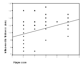
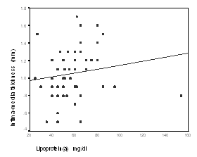
IntroductionLipoprotein(a) (Lp(a)) is a cholesterol-rich particle existing in human plasma, was first described by Berg in 1963 (1, 2). Many epidemiological and case-control studies have shown that, when present in high levels in plasma, Lp(a) is recognized as an independent risk factor for premature atherosclerotic coronary heart disease (1-3). The exact mechanism by which Lp(a) is a cardiovascular risk, is unknown, however, both proatherogenic and prothrombogenic effects have been hypothesized, however, the biological role and normal metabolism of this lipoprotein are not fully elucidated (1-3). In renal failure, studies revealed an increase in plasma concentration of Lp(a) (5,6). Elevated plasma Lp(a) levels in chronic renal failure patients have been associated with a frequency distribution of apolipoprotein(a) [apo(a)] isoforms, Similar to those found in general population. This indicates that elevated Lp(a) levels in these patients are not due to genetic origin (6,7) therefore, it has been suggested that kidneys have an important role in Lp(a) metabolism, decrease Lp(a) catabolism or increase of liver production (7-11). In fact, with beginning of chronic renal failure and as glumerular filtration rate (GFR) reach below 70ml/min, Lp (a) start to increase(11-13), and dialysis procedure by itself doesn’t seem to able to decrease the Lp(a) levels (5-8, 12). Irrespective of pathophysiological mechanisms involved, increased Lp(a) levels could be a contributing factor in the increased incidence of atherosclerotic disease observed in hemodialysis patients (12-18).The early stages of atherosclerosis are associated with changes in arterial structure. Subtle structural changes, such as thickening of arterial intimae-media complex (IMT), occur early in the atherosclerotic disease process (15-18). Using B-mode ultrasonography for assessing early arteriosclerosis is safe and non-invasive to study superficial vascular districts, such as the carotid and femoral arteries (15-19). Therefore ultrasonic evaluation of carotid artery for IMT and plaques can identify patients at risk for cardiovascular disease (15-18). Carotid arteries are privileged area for studying the progression of atherosclerotic lesions from onset to fully developed plaque. Carotid - IMT measurements are strongly related to the extent of atherosclerosis in other vascular districts too (15-18). As many known and conventional risk factors have been shown to be significantly associated with increased arterial wall thickness, consistent with their accepted role in atherogenesis, much less is known, however, about the effects of Lp(a) on IMT in patients under regular hemodialysis (1, 18), Therefore the aim of present study was to examine the effects of plasma Lp(a) levels on early structural atherosclerotic vascular changes in a group of end-stage renal failure patients under regular hemodialysis. Patients and MethodsThis is a cross-sectional study, was done on 61 unselected patients with end-stage renal disease (ESRD), undergoing maintenance hemodialysis treatment between September 2002 and decem¬ber2003.On patients selection exclusion criteria were cigarette smoking, body mass index (BMI) more than 25, anti lipid drugs taking, recent MI and vascular diseases as well as active or chronic infection and inclusion criteria was hypertention. For all subjects lipoprotein (a) measured by enzyme immunoassay (ELISA) by Immuno-Biological laboratories (IBL) kit , carotid sonography was done by a single sonologist unaware of history or lab data of patients, using a Honda-Hs-2000 Sonograph and 7.5 MHZ linear probe, IMTs in mm were measured, the procedure was done at the end of diastolic phase, the sites of measurements were at the distal common carotid artery, area of bifurcation and at the first proximal internal carotid artery, IMT was measured at the plaque free areas, for examination, subjects were in supine position with neck hyperextension and rotation of head for facilitation of procedure performing. Carotid evaluated in axial longitude. By sonography, the carotid artery found to has tree different echoes, Intimae, is as an echogen layer line, media hypoecho, and adventitia is echogen. Intimae- media thickness (IMT) was defined as the distance from leading edge of lumen-Intimae interface of the far wall to the leading edge of the media-adventitia interface of the far wall. IMT more than 0.8 mm was considered abnormal, for statistical analysis we measured the mean of right and left carotid IMT. Sonography for plaque was done at the right and left of carotid and femoral arteries and scored from 0 (no plaque) to score 4 (plaque presence at all four sites) regardless of the number and size of the plaques in each site, plaque occurrence in each site, scored one point, plaques divided into 3 groups, soft, calcified and mixed, plaques visualized as a echogen or hypoecho protrusion into the vessel lumen. Soft plaques are hypoecho, calcified are echogen and had shadow. Mixed plaques have heterogen echo, plaques was considered as a local intimal thickness more than 1 mm for plaque measurement the largest longitude was considered. For statistical analysis descriptive data are expressed as Mean± SD, comparison between groups were evaluated by using chi-square (χ2 test), Kruskal&Wallis test, Mann-Whitney U test and Fisher's exact test, for correlations we used Spearman test, partial correlation test with adjustment for age, Phi&Cramer's V test and Etta test. All statistical analysis were performed by using the statistical analysis system (SPSS version 11.00), statistical significance was inferred at a p value< 0.05. ResultsTotal patients were 61 (F=23 M=38), consisted of 50 non diabetic hemodialysis patients (F=20 M=30), and 11 diabetic hemodialysis patients (F=3 M=8), table 1 show the mean± SD of data, table 2 show the frequency distribution of plaque score in total patients consisted of diabetic and non diabetic groups (all of the plaques were calcified) and all of the hemodialysis patients were hypertensive from stage one to three. Mean±SD of ages of subjects were 46.5±16 years. Mean±SD of length of the time patients had been on hemodialysis were 32 ± 31 months. Mean±SD of LP(a) of total patients were 58.5±19mg/dl. Mean±SD of LP(a) of diabetic group were 62±12.3 mg/dl and for no diabetic group were 57.7±20 mg/dl. In this study there were no significant difference of ages, and duration of hemodialysis between males and females (p>0.05) (Mann-Whitney U test), no significant difference of DM between two sexes (Fisher's exact test) (p>0.05), no significant difference of duration of hemodialysis and age of the patients between diabetic and non diabetic group were existed (P>0.05) (Mann-Whitney U test). There was a significant different of carotid-IMT between diabetic and no diabetic group (1.3±0.3 versus 1±0.25 mm respectively) (p<0.05) (Etta test). No significant difference of duration of hemodialysis treatment as well as serum lipoprorein(a) and ages of patients between diabetic and non diabetic group (p>0.05) (Mann-Whitney U test) were existed, about plaque score, positive correlation of plaque scores with ages of the patients (p=0.003), was demonstrated (Kruskal-Wallis test). Significant positive correlation between plaque scores and diabetes mellitus was observed (p=0.004) (χ2 test). Significant positive correlation between plaque score with length of the time patients had been on hemodialysis (r=0.239 p=0.033) as well as significant positive correlation between Carotid-Femoral plaques (plaque scores) with Carotid- IMT was demonstrated (r=0.306 p=0.009) (partial correlation test after adjustment for age) (figure1). Statistical analysis on Carotid-IMT with partial correlation test (after adjustment for ages) showed no positive correlation between IMT with duration of hemodialysis (p>0.05). Statistical analysis on LP(a) showed significant positive correlation between Carotid-IMT with lipoprotein(a) (r= 0.330 p=0.009) (figure2) (Spearman test), also there was a significant positive correlation between plaque score with lipoprotein(a) (r=0.205 p=0.051) (figure3) (partial correlation test after adjustment for age). Finally after divided of patients into diabetic and non diabetic for analysis of the correlation mentioned above, in two groups respectively, no other important data were found. DiscussionIn this study there were, more thickening of Intima-media complex in diabetic group, positive association of plaque score with ages and DM, positive correlation of carotid-IMT with carotid-femoral plaque score, positive corration of serum LP(a) with carotid-IMT and carotid-femoral plaque score, no significant difference of LP(a) between diabetic and non diabetic HD patients. Pascasio et al. observed a large number of vascular plaques in uremia patients, he concluded that the process of advance atherosclerosis might be started with the beginning of renal failure, he suggested that hemodialysis treatment may not a potential factor to accelerate arthrosclerosis finally he concluded, the progression of atherosclerosis might be related to atherogenic factors operative before regular dialysis (19). Damjanovic et al. evaluated IMT of 45 dialysis patients, found higher mean carotid IMT in HD patients than in control group, he showed positive correlation of IMT with certain risk factors for atherosclerosis (age, duration of dialysis and lipid parameters) (20). Correlation of IMT with ages and duration of hemodialysis in HD patients was evaluated, by Shoji and Hojs et al. no clear relationship of IMT with duration of hemodialysis were found in their studies (21,22). Hojs also in his study (on 28 HD patients) observed, age was the only significant determinant of number of plaques, he concluded that hemodialysis patients had advanced atherosclerosis in the carotid arteries compared with normal subjects (22). More over Hojs in his recent study, showed no difference in plaque occurrence between 28 hemodialusis patients with 28 ESRD patients prior to hemodialysis (23). Savage et al. in a study on 24 dialysis patients noted on more prevalence of plaque in carotid and femoral artery. Also this study showed the relationship between femoral artery plaque and age also he reported the correlation of age with IMT of carotid artery of HD patients (24). Moreover in a recent study by Kato A et al. showed a significant correlation of IMT with age on 219 HD patients(25). Sramek A. et al. on 142 asymptomatic men, found no increased IMT in the carotid or femoral artery at high levels of Lp (a), he concluded that, Lp(a) levels are not associated with early atherosclerotic vessel wall changes in the carotid or femoral arteries (26). Dentil et al. in a study on 100 elderly subjects (aged 78.5±0.6), showed no association between carotid IMT and Lp (a), he concluded, Lp (a) was unrelated to the severity of extra cranial vessels atherosclerosis (27), while Baldassarre D et al in a study on 100 type 2 hypercholestrolemic patients, showed higher values of carotid IMT in hyrercholestrolemic patients with plasma Lp (a) levels > 30 mg/dl than in those with lower levels, he concluded, elevated plasma levels of Lp (a) can be considered an additional independent factor associated with thickening of carotid ar¬tery in patients with severe hypercholesterolemia but not in those with moderate hypercholesterolemia or normocholestrolemic subjects (28). Finally Raitakari et al. on 241 healthy subjects suggested no association between IMT and Lp(a) but significant positive correlation with total Cholesterol, LDL-c, LDL/HDL ratio, age, and Tg were found (1). In the present study, a significant association between serum lipoprotein(a) with carotid-IMT and carotid-femoral plaques were found, as the extraordinary high mortality in end-stage-renal disease (ESRD) patients under hemodialysis are due to cardiovascular disease, there is some interest to non traditional atherosclerotic cardiovascular disease risk factors that are Prevalent in ESRD, Such as Lipoprotein(a), needs to more attention, because of its effect to acceleration of rapid progressive atherosclerosis seen in HD patients.
|
||||||||||||||||||||||||||||||||||||||||||||||||||||||||||||||
*duration of hemodialysis treatment |
||||||||||||||||||||||||||||||||||||||||||||||||||||||||||||||
 |
 |
|||||||||||||||||||||||||||||||||||||||||||||||||||||||||||||
|
Figure1: Significant positive correlation of Carotid-Femoral-plaque score with Carotid-IMT (r= 0.306 p=0.009) (partial correlation test after adjustment for age). |
Figure 2: Significant positive correlation of Carotid-IMT with LP(A) ( r= 0.330 p=0.009) (Spearman test) . |
|||||||||||||||||||||||||||||||||||||||||||||||||||||||||||||
|
Table2: Frequency distribution of plaque scores of Carotid and Femoral arteries. |
||||||||||||||||||||||||||||||||||||||||||||||||||||||||||||||
Group3=No diabetic HD patients |
||||||||||||||||||||||||||||||||||||||||||||||||||||||||||||||
AcknowledgmentWe would like to thank DR. M. Mowlaie (sonologist) for carotid ultrasonographies.. References
Corresponding Address: |
||||||||||||||||||||||||||||||||||||||||||||||||||||||||||||||
|
|
||||||||||||||||||||||||||||||||||||||||||||||||||||||||||||||
| Infotrend Crea(c)tive Design | Revised: 20 May 2009 | |||||||||||||||||||||||||||||||||||||||||||||||||||||||||||||
