|
||||||||||||||||||||||||||||||||||||
| [
Contents
] [ INDEX ]
|
||||||||||||||||||||||||||||||||||||
|
Page 89 |
||||||||||||||||||||||||||||||||||||
|
Case report Foreign bodies in the urinary bladder - case report (1) Radoš Žikić, (2) Zoran Jelenković, (3) Zvonimir Adamović, (3) Vladan Radojević (1) POLYCLINIC "PAUNKOVIĆ" ZAJEČAR; (3) HEALTH CENTER ZAJEČAR, ZAJEČAR |
||||||||||||||||||||||||||||||||||||
|
|
||||||||||||||||||||||||||||||||||||
| Download in pdf format | Abstract:
INTRODUCTION Pathological substances produced by the body and
entering the urinary bladder cannot be considered foreign bodies.
They are more common in women than in men, with a ratio of 100:1
according to some statistics (Sonntag). They can be of animal,
plant, or mineral origin. Medical foreign bodies remain after
certain surgical interventions, either in the bladder itself or on
surrounding organs, due to negligence, carelessness, incorrect use,
or poor quality of materials. CASE REPORT Patient C.R. from the
vicinity of Donji Milanovac, aged 30, presented to our clinic
complaining of frequent urination, burning sensation during
urination, and occasional sudden cessation of urination. Anamnestic
data were very scarce, except for the mentioned symptoms. Urine
sediment showed a significant number of pale red blood cells and
rare bacteria. Urine culture yielded sterile cultures. Descending
cystography revealed an ellipsoid contrast defect. Subsequent
urethrocystoscopy showed a urethra easily passable for a Ch 20
cystoscope sheath, with a short prostatic urethra of about 2 cm.
CONCLUSION Operative findings revealed: A round dark-brown
formation, the size of a pigeon's egg, was observed in the bladder
lumen. A "stone" was extracted from the bladder lumen with stone
forceps and placed in a kidney basin. After completing the
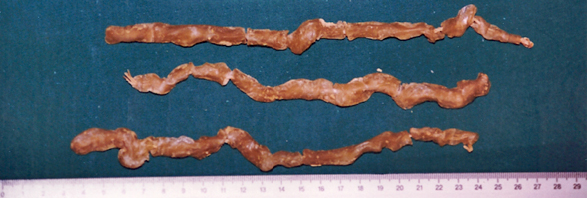
operation, the image showed a foreign body. Three snake-like
objects, twisted, with a length of about 10 cm and a thickness of
about 10 mm, were found in the kidney basin. They were candles. Keywords: urinary bladder foreign body, Cystoscopy, candles, quackery |
|||||||||||||||||||||||||||||||||||
|
INTRODUCTION Pathological substances produced by the body and entering the
urinary bladder cannot be considered foreign bodies. They are more
common in women than in men, with a ratio of 100:1 according to some
statistics (Sonntag). They can be of animal, plant, or mineral
origin. Medical foreign bodies remain after certain surgical
interventions, either in the bladder itself or on surrounding
organs, due to negligence, carelessness, incorrect use, or poor
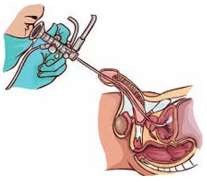
quality of materials. Patient C.R. from the vicinity of Donji Milanovac, aged 30, presented to our clinic complaining of frequent urination, burning sensation during urination, and occasional sudden interruption of urination. Anamnestic data are very scarce, except for the mentioned symptoms. Urine sediment analysis revealed numerous pale red blood cells and occasional bacteria. Urine culture showed sterile growth media. Descending cystography revealed an ellipsoid defect in contrast. Subsequently, urethrocystoscopy was performed (Figure 1): the urethra allowed easy passage for the sheath of the 20 Fr cystoscope, with the prostatic urethra measuring approximately 2 cm in length. Figure 1. Urethrocystoscopy Cystoscopy (taken from: http://cdn.futura-sciences.us/builds/images/thumbs/4/41b85c2a84_cystoscopie-c-hakan-corbac-305-fotoliacom.jpg)
The bladder neck is normal. A round dark-brown formation, the
size of a pigeon egg, is observed within the bladder lumen.
Suspecting a calculus, attempts were made to perform
electrohydraulic lithotripsy with Urat-I. However, stone
disintegration was unsuccessful, leading to a recommendation for
surgical stone removal. Figure 2. Foreign bodies - candles, surgically removed from the bladder
To our astonishment, three snake-like objects, curved,
approximately 10 cm in length and 10 mm in diameter, were found in
the kidney. They turned out to be candles. DISCUSSION In most cases, foreign bodies in the urinary bladder occur in
individuals with psychopathological, mentally impaired, or
intoxicated states. Small children, in innocent play, often insert
various objects into natural orifices. Women may accidentally or due
to negligence or lack of knowledge insert contraceptive devices or
abortion aids into the urethra instead of the vagina. Medical
foreign bodies may remain accidentally due to carelessness,
negligence, incorrect use, or poor material during a surgical
intervention in the bladder itself or on surrounding organs. Even
surgical suture materials can act as a nucleus around which a stone
forms. Firearm injuries, more common in war and rarer in peacetime,
can create a foreign body due to a retained projectile in the
bladder or its surroundings. Cases of projectile migration from the
abdomen into the urinary bladder have been described. CONCLUSION Patient C.R. from the vicinity of Donji Milanovac, aged 30,
presented to our clinic complaining of frequent urination, burning
sensation during urination, and occasional sudden interruption of
urination. On descending cystography, an elliptical contrast defect
was observed. Subsequently, urethrocystoscopy was performed,
revealing a urethra easily passable for the sheath of a size 20
cystoscope, with a prostatic urethra approximately 2 cm in length. LITERATURE:
|
||||||||||||||||||||||||||||||||||||
|
|
||||||||||||||||||||||||||||||||||||
| [
Contents
] [ INDEX ]
|
||||||||||||||||||||||||||||||||||||
|
||||||||||||||||||||||||||||||||||||