Glasilo Podružnice Srpskog lekarskog društva Zaječar

Godina 2019     Volumen 44     Broj 1
     
      [ Sadržaj ] [ Indeks autora ] <<< ] >>> ]      
      UDK 616.24-003.4
COBISS.SR-ID 276243212

ISSN 0350-2899. - Vol. 44, br. 1 (2019),
str. 22-25.


Prikaz slučaja / Case report

Large pulmonaly cyst in nonsmoker - sporadic finding
Velika plućna cista kod nepušača- sporadičan nalaz

Biljana Lazović (1), Mihajlo Korać (1), Vuk Aleksić (1), Branko Lukić (1), Dejan Stevanović (1), Damir Jašarović (1), Rade Milić (2), Vladimir Žugić (3)

(1) UNIVERSITY CLINICAL CENTER "ZEMUN", BELGRADE, SERBIA, (2) MILITARY MEDICAL ACADEMY, CLINIC FOR PULMONARY DISEASE, BELGRADE, SERBIA, (3) CLINIC FOR LUNG DISEASE, SCHOOL OF MEDICINE, UNIVERSITY OF BELGRADE, SERBIA


Preuzmite rad u pdf formatu

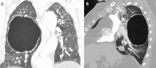
  Sažetak: Cistična bolest pluća (CLD) je grupa oboljenja pluća koju karakteriše prisustvo multiplih cisti, definisanih kao translucencije ispunjene vazduhom ili zone niske atenuacije, oivičene tankim zidom (uglavnom < 4mm). Često se mešaju sa džinovskim bulama koje su uobičajene kod pacijenata sa emfizemom. Prikaz slučaja: Žena stara 76 godina sa prethodnom medicinskom istorijom o subokluzivnim smetnjama primljena je na odeljenje hirurgije. Uzgredno, prilikom preoperativnog pregleda RTG snimka grudnog koša uočena je džinovska bula u desnom plućnom krilu. Kompjuterizovana tomografija je potvrdila nalaz, ali zbog svoje debljine zida promena je svrstana u plućne ciste koje imaju potpuno drugačiji pristup u lečenju. Zbog toga, pacijentkinja nije podvrgnuta operaciji i lečena je konzervativno. Zaključak: Glavna razlika između džinovskih bula i plućnih cisti je debljina zida, ukoliko je od 1 – 4 mm radi se o plućnoj cisti, za razliku od bule čiji je zid tanji od 1 mm.
Ključne reči: ciste, bule, HOBP, imidžing

Summary: Cystic lung disease (CLD) is a group of lung disorders characterized by the presence of multiple cysts, defined as air-filled lucencies or low-attenuating areas, bordered by a thin wall (usually < 4 mm). It is very often mixed with giant bullaes which are common in patients with emphysema.
Case report: A 76-year-old woman with a medical history of subocclusive disturbance was admitted to surgery department. Accidentally, during preoperative examination, chest X-ray revealed giant bullae in the right lung. CT scan confirmed it, but due to the wall thickness the changes have been classified into pulmonary cyst which have a completely different approach in treatment. Thus, patient underwent without surgery and was treated conservatively. Conclusion: The main difference between a giant bullae and a pulmonary cyst is in wall thickness, if it measures between 1 and 4 mm it is pulmonary cyst, unlike bullae whose wall is thinner than 1mm.
Key words: smokers, cystae, bullae, COPD, imaging


Introduction:

Cigarette smoking is the major risk factor for the development of chronic obstructive pulmonary disease (COPD), but not all smokers develop COPD, and such patients deserve a workup for one of the less common causes of emphysema. These conditions include A1AT deficiency, connective issue disease (Cutis laxa, Marfan syndrome, Ehler-Danlos syndrome), intravenous drug abuse (methylphenidate, cocaine or talc), HIV infection, hypocomplementemic urticarial vasculitis syndrome, malnutrition and several rare metabolic disorders (Salla disease, Menke syndrome).[1] Other differential diagnoses of bullous emphysema and bullous lung disease include uncommon causes such as autoimmune diseases (Sjögren disease, Wegener granulomatosis disease and multisystem autoimmune dysfunction), bullous sarcoidosis, Birt-Hogg-Dubé syndrome, neurofibromatosis, placental transmogrification of the lung, Fabry disease, idiopathic giant bullous emphysema, etc.[2] Almost all these conditions have systemic manifestation with characteristic features including early onset, liver dysfunction, vasculitis, skin and joint manifestations, lymphadenopathy, etc. The evaluation of bullous emphysema in nonsmokers therefore begins with a detailed medical history, including age of onset of the disease and a physical examination, including determination of the presence of extrapulmonary symptoms or signs and measurement of A1AT level.
On the other side, pulmonary cysts are cystic spaces within the lung parenchyma that have a wall that measures between 1 and 4 mm. Lung cysts contain gas, not fluid. The thickness of the wall is what distinguishes a cyst from a pulmonary tissue (that is defined as having a wall that is thicker than 4 mm). A bulla has a wall that is less than 1 mm. In contradistinction to all other organs, the term cyst as used in the lung, is a misnomer, as it refers to a contained focus of gas, not fluid. Pulmonary cysts can be congenital or acquired [1, 2]. Incidental findings of pulmonary cysts are becoming more common because of the widespread use of CT scans in daily clinical practice and in lung cancer screening. Multiple pulmonary cysts are identified in various diseases such as pulmonary Langerhans cell histiocytosis (PLCH), lymphangioleiomyomatosis (LAM), and lymphoid interstitial pneumonia (LIP) [3, 5]. These progressive diseases are often symptomatic and may result in impairment of pulmonary functions. In contrast, solitary or sporadic pulmonary cysts can be incidentally seen on chest CT of otherwise healthy individuals [5, 6].
Pulmonary bullae are focal regions of emphysema with no discernible wall which measure more than 1 cm in diameter which makes difference from pulmonaly cyst. Sometimes it is very difficult to distinguish these two identities [1].
We present a 76 years old female who was admitted to surgery clinic due to subocclusive interference, confirmed radiologically, for detailed examinations. She is a housewife, living on a mountain at 1500 meters above sea level whole her life. She had no other comorbidities. Abdomen pain started 7 days before admission and sometimes it was extremely painful. NCT scan of abdomen showed no pathological findings. She was preparing for laparoscopy surgery and surgeon asked for chest X ray, which described a large bullae measuring 111x88x75 mm in diameter. Pulmonologist asked for CT of thorax, which confirmed a large bullae - differential diagnosis pulmonary cyst. Detailed analysis confirmed pulmonary cyst according the guidelines (Figure 1A, B). Spirometric tests showed reduced FVC and FEV1 with a reduced FEV1/FVC ratio suggesting mild airways obstruction. Physical examination was unremarkable. Measurements of A1AT level were normal. Laboratory and biochemical analyses were in physiological range.
In the meantime, her abdomen disturbances were passed on to a conservative treatment and she was discharged from hospital.

Discussion:

The harmful effects of tobacco smoking, especially with regard to the aetiology of chronic obstructive pulmonary disease, are well documented [1, 2]. Large emphysematous bullae
could be developped, usually in the context of significant tobacco exposure over many years, and tend to be associated with airway obstruction, reduced gas transfer factor and diffusion coefficient, and evidence of centrilobular emphysema elsewhere in the lungs [7]. On the other hand, clinical awareness of cystic lung disease (CLD) has recently increased due to the widespread use of high-resolution computed tomography HRCT) and the country’s general-health medical examinations [8]. Cysts are rare in asymptomatic individuals < 55 years of age but their prevalence increases with age [9]. CLD is a constellation of diverse lung disorders that originate from various causes but they have in common an anatomical cystic deformity. Due to its etiological heterogeneity, CLD can be classified based on the underlying pathophysiologic mechanisms: congenital, genetic, infectious, inflammatory, lymphoproliferative, neoplastic, and smoking-related [10,11]. CLD must also be distinguished from emphysema, cavity, bulla, pneumatocele, and honeycombing, all of which feature lucencies that mimic a pulmonary cyst. These can be differentiated from pulmonary cysts based on the thickness of the wall, the size of the gas-filled space, the anatomic location, and the tendency of true cysts to cluster [9].

Figure 1. A 76-year old female patient with pulmonary cyst. At CT a giant air filled pulmonary cyst occupying apical segment of right lower lobe (111x88x75 mm; CCxLLxAP) lined with regular wall thickness around 2mm, partially calcified, without communication with the bronchial tree and without compression of adjacent lung tissue. That is consistent with a chronic state, most probable a congenital lesion.
Slika 1: 76-godišnja bolesnica sa plućnom cistom. Na CT-u se nalazi ogromna vazdušna plućna cista koja zauzima apikalni segment desnog donjeg režnja (111x88x75 mm; CCxLLxAP) obložen pravilnim zidom debljine oko 2mm, delimično kalcifikovan, bez komunikacije sa bronhijalnim stablom i bez kompresije susednog plućnog tkiva, što je u skladu sa hroničnim stanjem, najverovatnije kongenitalnom lezijom.

Conclusion:

With the increased use of CT, the recognition of CLD has also increased. HRCT is the most valuable diagnostic modality for an initial evaluation of CLD as well as bullaes. Along with the characteristic findings of cysts on CT, the pathologic features, presence of genetic mutation, and concomitant disease may be helpful in establishing the differential diagnosis. However, as CLDs are rare, treatment is currently insufficient. Therefore, enhanced knowledge of the pathogenesis of CLDs is needed to guide therapeutic decision-making for each entity.
Zaključak:
Sa povećanom upotrebom CT-a, prepoznavanje cistične bolesti pluća (CLD) se takođe povećalo. HRCT je najvredniji dijagnostički modalitet za početnu procenu kako CLD-a tako i bula. Karakteristični nalazi cista na CT, uz patološka svojstva, prisustvo genetske mutacije i pratećih bolesti mogu biti od pomoći u uspostavljanju diferencijalne dijagnoze. Međutim, kako su CLD retki, lečenje je trenutno nedovoljno. Stoga je potrebno bolje poznavanje patogeneze CLD-a koje bi uticalo na terapijsko donošenje odluka za svaki entitet.

REFERENCES:

  1. Lee P, Gildea TR, Stoller JK. Emphysema in nonsmokers: Alpha 1-antitrypsin deficiency and other causes. Cleveland Clin J Med. 2002;69: 928–46.
  2. Teramoto S, Fukuchi Y. Bullous emphysema. Curr Opin Pulm Med. 1996;2:90–6
  3. Priest JR, Williams GM, Hill DA et al. Pulmonary cysts in early childhood and the risk of malignancy. Pediatr. Pulmonol. 2009;44 (1): 14-30
  4. Cantin L, Bankier AA, Eisenberg RL. Multiple cystlike lung lesions in the adult. AJR Am J Roentgenol. 2010;194 (1):W1-W11
  5. Hansell DM, Bankier AA, MacMahon H, McLoud TC, Müller NL, Remy J. Fleischner Society: Glossary of Terms for Thoracic Imaging. Radiology. 2008; 246(3): 697–722.
  6. Elicker BM, Webb WR. Fundamentals of High-Resolution Lung CT: Common Findings, Common Patterns, Common Diseases and Differential Diagnosis. Lippincott Williams & Wilkins; 2013.
  7. Tashkin DP, Coulson AH, Clark VA, Simmons M, Bourque LB, Duann S, Spivey GH, et al. Respiratory symptoms and lung function in habitual heavy smokers of marijuana alone, smokers of marijuana and tobacco, smokers of tobacco alone and nonsmokers. Am Rev Respir Dis. 1987; 135:209–16.
  8. Park HY, Nam HS, Chung MP, et al. A nationwide survey of lymphangioleiomyomatosis in Korea: recent increase in newly diagnosed patients. J Korean Med Sci. 2010;25:1182–1186
  9. Copley SJ, Wells AU, Hawtin KE, Gibson DJ, Hodson JM, Jacques AE, et al. Lung morphology in the elderly: comparative CT study of subjects over 75 years old versus those under 55 years old. Radiology. 2009; 251(2):566–73
  10. Gupta N, Vassallo R, Wikenheiser-Brokamp KA, McCormack FX. Diffuse cystic lung disease: part I. Am J Respir Crit Care Med. 2015; 191:1354–1366
  11. Kwon YS, Han J, Jung KH, Kim JH, Koh WJ. Mycobacterium avium lung disease combined with a bronchogenic cyst in an immunocompetent young adult. Korean J Intern Med. 2013; 28:94–97


      Adresa autora / Corresponding address:
Biljana Lazović,
University clinical center "Zemun", Vukova 9, Zemun, 11070 Belgrade
E-mail: lazovic.biljana@gmail.com
Rad primljen: 28.2.2019.
Elektronska verzija objavljena: 13.5.2019.


      [ Sadržaj ] [ Indeks autora ] <<< ] >>> ]      


Timočki medicinski glasnik, Zdravstveni centar Zaječar
Journal of Regional section of Serbian medical association in Zajecar
Rasadnička bb, 19000 Zaječar, Srbija
E-mail: tmglasnik@gmail.com

Pretraživanje / Site Search

  www.tmg.org.rs


      Design: Infotrend  
         

counter on myspace